Fosi S, Giuricin V, Girardi V, Di Caprera E, Costanzo E, Di Trapano R, Simonetti G
Department of Diagnostic Imaging, Molecular Imaging, Interventional Radiology and Radiation Therapy, University Hospital Tor Vergata, Viale Oxford 81, 00133 Rome, Italy.
Case Rep Radiol. 2014;2014:431563. doi: 10.1155/2014/431563. Epub 2014 Jul 17.
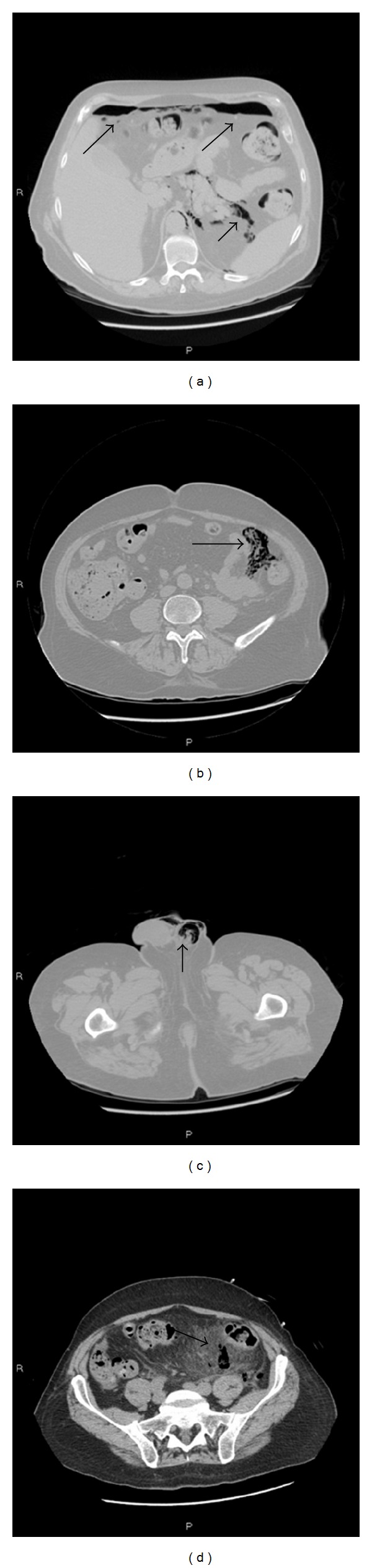
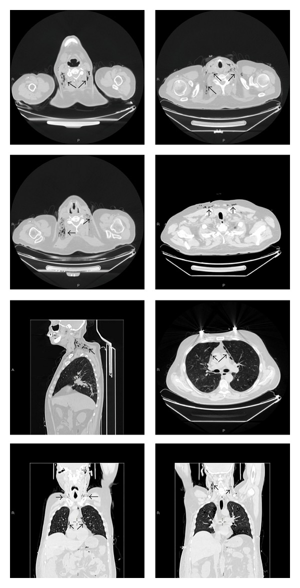
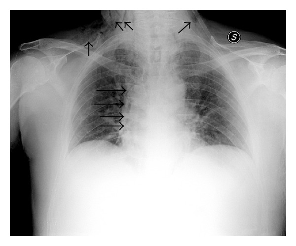
Pneumomediastinum, and subcutaneous emphysema usually result from spontaneous alveolar wall rupture and, far less commonly, from disruption of the upper airways or gastrointestinal tract. Subcutaneous neck emphysema, pneumomediastinum, and retropneumoperitoneum caused by nontraumatic perforations of the colon have been infrequently reported. The main symptoms of spontaneous subcutaneous emphysema are swelling and crepitus over the involved site; further clinical findings in case of subcutaneous cervical and mediastinal emphysema can be neck and chest pain and dyspnea. Radiological imaging plays an important role to achieve the correct diagnosis and extension of the disease. We present a quite rare case of spontaneous subcutaneous cervical emphysema, pneumomediastinum, and pneumoretroperitoneum due to perforation of an occult sigmoid diverticulum. Abdomen ultrasound, chest X-rays, and computer tomography (CT) were performed to evaluate the free gas extension and to identify potential sources of extravasating gas. Radiological diagnosis was confirmed by the subsequent surgical exploration.
纵隔气肿和皮下气肿通常由肺泡壁自发性破裂引起,而上呼吸道或胃肠道破裂导致的情况则极为罕见。结肠非创伤性穿孔引起的颈部皮下气肿、纵隔气肿和腹膜后气肿鲜有报道。自发性皮下气肿的主要症状是受累部位肿胀和捻发音;颈部和纵隔皮下气肿的进一步临床发现可能为颈部和胸部疼痛以及呼吸困难。放射影像学在正确诊断疾病及其范围方面起着重要作用。我们报告一例极为罕见的因隐匿性乙状结肠憩室穿孔导致的自发性颈部皮下气肿、纵隔气肿和腹膜后气肿病例。进行腹部超声、胸部X线和计算机断层扫描(CT)以评估游离气体的范围并确定气体外渗的潜在来源。后续手术探查证实了放射学诊断。