Zhang Jiong, Yanez David, Floege Anna, Lichtnekert Julia, Krofft Ronald D, Liu Zhi-Hong, Pippin Jeffrey W, Shankland Stuart J
Division of Nephrology, Department of Medicine, University of Washington School of Medicine, Washington Current address: Research Institute of Nephrology, Jinling Hospital, Nanjing University School of Medicine, China.
Department of Biostatistics, School of Public Health, University of Washington, Washington.
J Renin Angiotensin Aldosterone Syst. 2015 Jun;16(2):234-48. doi: 10.1177/1470320314543910. Epub 2014 Aug 20.
The objective of this article is to test the effects of angiotensin-converting enzyme (ACE)-inhibition on glomerular epithelial cell number in an inducible experimental model of focal segmental glomerulosclerosis (FSGS).
Although ACE-inhibition has been shown to limit podocyte loss by enhancing survival, little is known about its effect on podocyte number following an abrupt decline in disease.
Experimental FSGS was induced with cytotoxic antipodocyte antibody. Following induction, groups were randomized to receive the ACE-inhibitor enalapril, the smooth muscle relaxant hydralazine (blood pressure control) or drinking water. Blood pressure, kidney function and histology were measured seven and 14 days following disease induction.
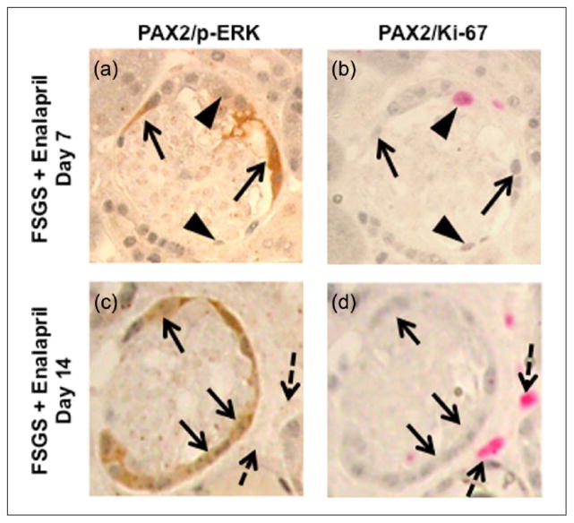
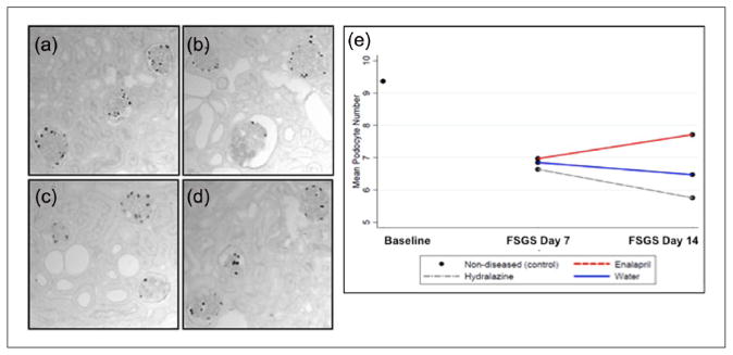
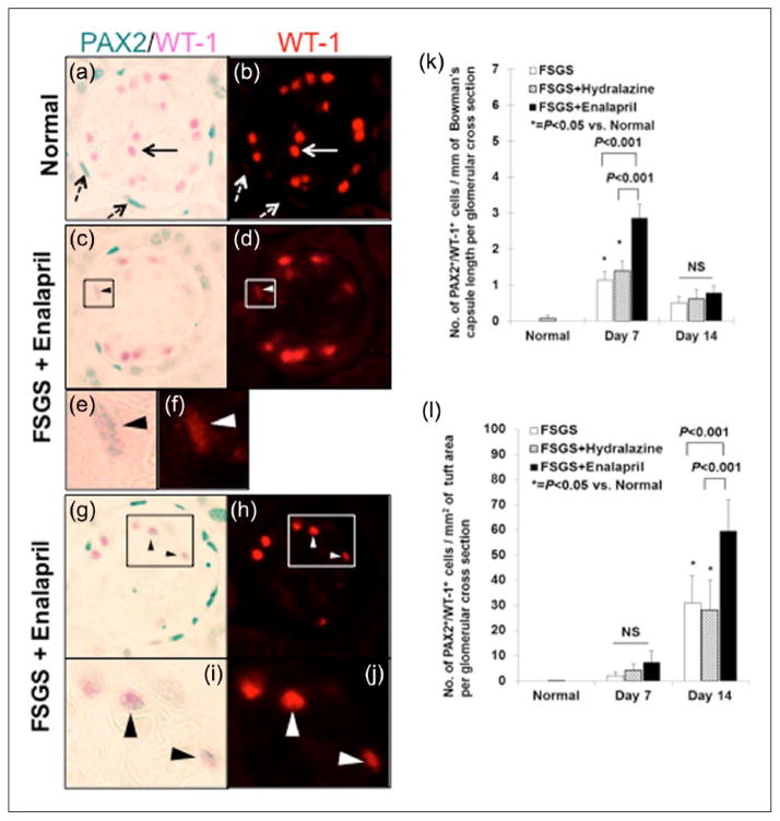
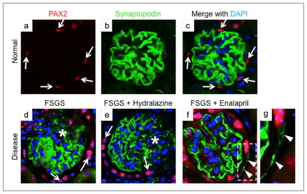
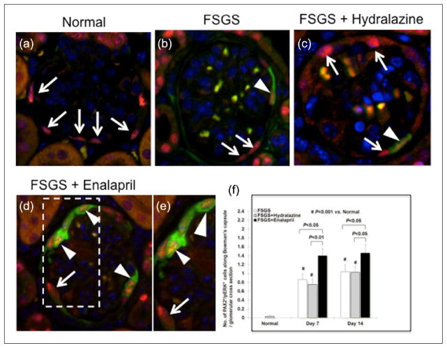
Both glomerulosclerosis and urinary albumin-to-creatinine ratio were less in the ACE-inhibition arm at day 14. At day 7 of disease, mean podocyte numbers were 26% and 29% lower in the enalapril and hydralazine arms, respectively, compared to normal mice in which no antibody was injected. At day 14, the mean podocyte number was only 18% lower in the enalapril arm, but was 39% lower in the hydralazine arm compared to normal mice. Podocyte proliferation did not occur at any time in any group. Compared to water- or hydralazine-treated mice with FSGS, the enalapril arm had a higher mean number of glomerular parietal epithelial cells that co-expressed the podocyte proteins WT-1 and synaptopodin, as well as phospho-ERK.
The results show following an abrupt decline in podocyte number, the initiation of ACE-inhibition but not hydralazine, was accompanied by higher podocyte number in the absence of proliferation. This was accompanied by a higher number of parietal epithelial cells that co-express podocyte proteins. Increasing podocyte number appears to be accompanied by reduced glomerulosclerosis.
本文旨在检测血管紧张素转换酶(ACE)抑制剂对局灶节段性肾小球硬化(FSGS)诱导实验模型中肾小球上皮细胞数量的影响。
尽管已证明ACE抑制剂可通过提高存活率来限制足细胞丢失,但对于疾病突然进展后其对足细胞数量的影响知之甚少。
用细胞毒性抗足细胞抗体诱导实验性FSGS。诱导后,将各组随机分为接受ACE抑制剂依那普利、平滑肌松弛剂肼屈嗪(血压控制)或饮用水组。在疾病诱导后7天和14天测量血压、肾功能和组织学。
在第14天,ACE抑制剂组的肾小球硬化和尿白蛋白与肌酐比值均较低。在疾病第7天,依那普利组和肼屈嗪组的平均足细胞数量分别比未注射抗体的正常小鼠低26%和29%。在第14天,依那普利组的平均足细胞数量仅比正常小鼠低18%,但肼屈嗪组比正常小鼠低39%。在任何组中,任何时候均未发生足细胞增殖。与FSGS的水或肼屈嗪治疗小鼠相比,依那普利组共表达足细胞蛋白WT-1和突触素以及磷酸化ERK的肾小球壁层上皮细胞平均数量更高。
结果表明,在足细胞数量突然下降后,开始使用ACE抑制剂而非肼屈嗪,在无增殖的情况下伴随着更高的足细胞数量。这伴随着共表达足细胞蛋白的壁层上皮细胞数量增加。足细胞数量增加似乎伴随着肾小球硬化减轻。