Roeder Sebastian S, Barnes Taylor J, Lee Jonathan S, Kato India, Eng Diana G, Kaverina Natalya V, Sunseri Maria W, Daniel Christoph, Amann Kerstin, Pippin Jeffrey W, Shankland Stuart J
Division of Nephrology, University of Washington, Seattle, Washington, USA; Friedrich-Alexander-Universität Erlangen-Nürnberg (FAU), Erlangen, Germany.
Division of Nephrology, University of Washington, Seattle, Washington, USA; Department of Biology, Oregon State University, Corvallis, Oregon, USA.
Kidney Int. 2017 Apr;91(4):896-913. doi: 10.1016/j.kint.2016.10.015. Epub 2016 Dec 18.
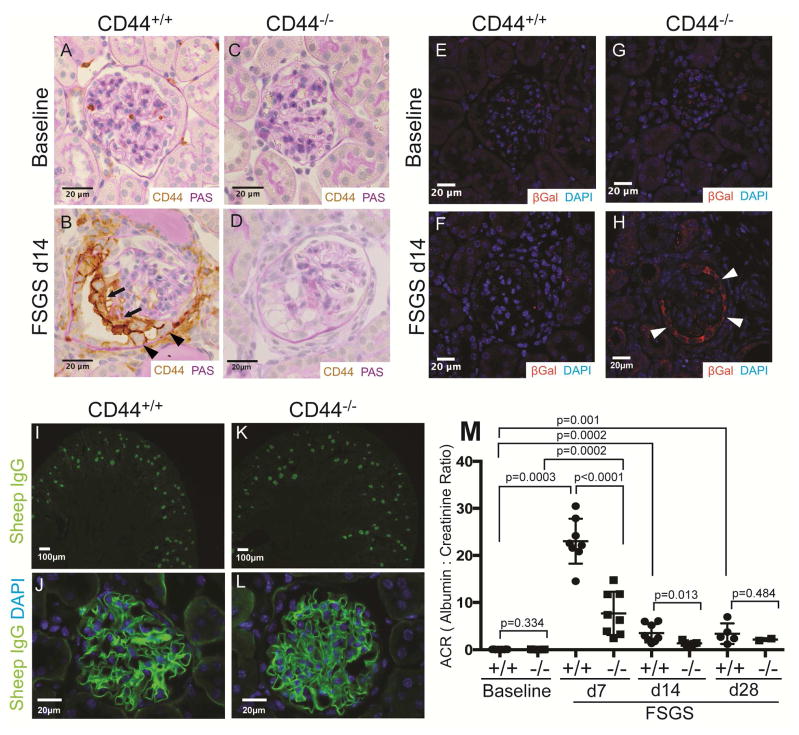
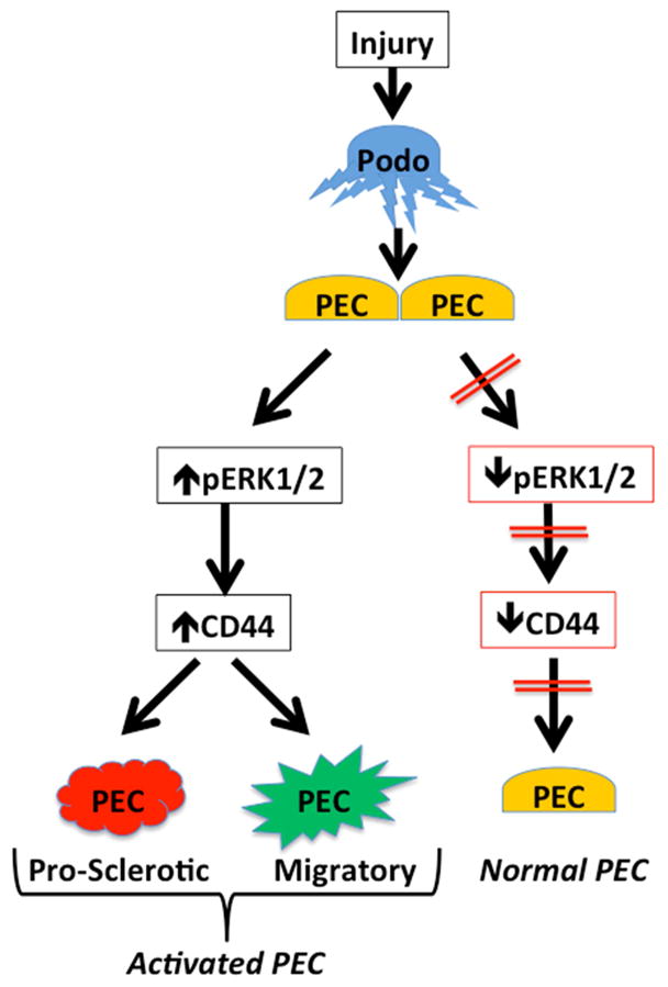
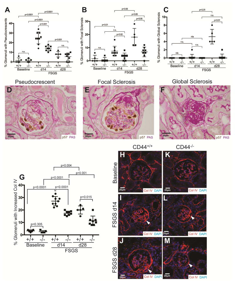
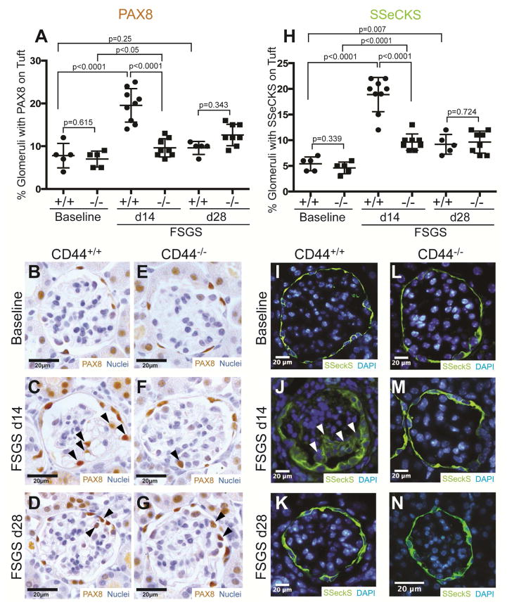
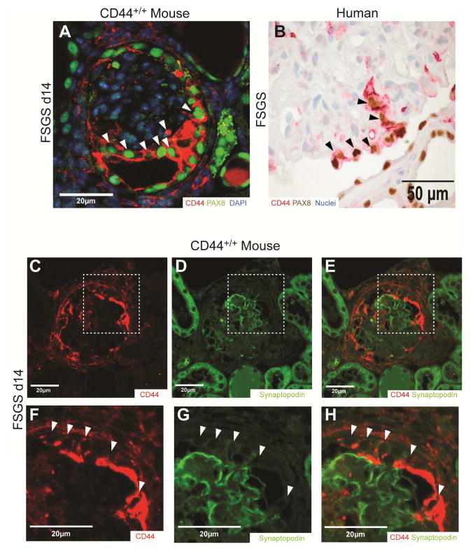
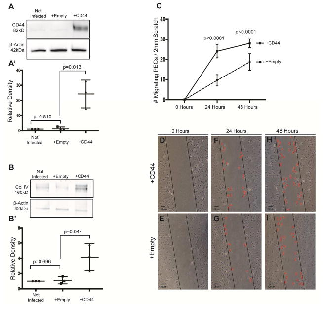
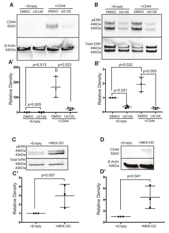
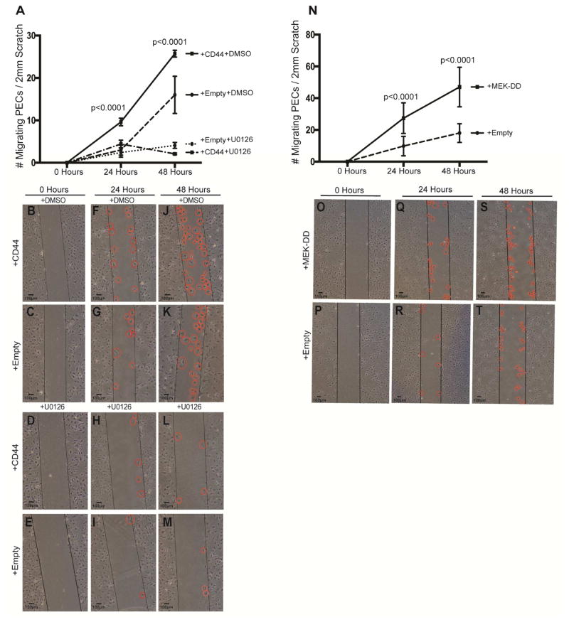
The glycoprotein CD44 is barely detected in normal mouse and human glomeruli, but is increased in glomerular parietal epithelial cells following podocyte injury in focal segmental glomerulosclerosis (FSGS). To determine the biological role and regulation of CD44 in these cells, we employed an in vivo and in vitro approach. Experimental FSGS was induced in CD44 knockout and wild-type mice with a cytotoxic podocyte antibody. Albuminuria, focal and global glomerulosclerosis (periodic acid-Schiff stain), and collagen IV staining were lower in CD44 knockout compared with wild-type mice with FSGS. Parietal epithelial cells had lower migration from Bowman's capsule to the glomerular tuft in CD44 knockout mice with disease compared with wild type mice. In cultured murine parietal epithelial cells, overexpressing CD44 with a retroviral vector encoding CD44 was accompanied by significantly increased collagen IV expression and parietal epithelial cell migration. Because our results showed de novo co-staining for activated ERK1/2 (pERK) in parietal epithelial cells in experimental FSGS, and also in biopsies from patients with FSGS, two in vitro strategies were employed to prove that pERK regulated CD44 levels. First, mouse parietal epithelial cells were infected with a retroviral vector for the upstream kinase MEK-DD to increase pERK, which was accompanied by increased CD44 levels. Second, in CD44-overexpressing parietal epithelial cells, decreasing pERK with U0126 was accompanied by reduced CD44. Finally, parietal epithelial cell migration was higher in cells with increased and reduced in cells with decreased pERK. Thus, pERK is a regulator of CD44 expression, and increased CD44 expression leads to a pro-sclerotic and migratory parietal epithelial cell phenotype.
糖蛋白CD44在正常小鼠和人类肾小球中几乎检测不到,但在局灶节段性肾小球硬化症(FSGS)中足细胞损伤后,肾小球壁层上皮细胞中CD44会增加。为了确定CD44在这些细胞中的生物学作用及其调控机制,我们采用了体内和体外研究方法。用细胞毒性足细胞抗体在CD44基因敲除小鼠和野生型小鼠中诱导实验性FSGS。与患有FSGS的野生型小鼠相比,CD44基因敲除小鼠的蛋白尿、局灶性和弥漫性肾小球硬化(过碘酸希夫染色)以及IV型胶原染色水平更低。与野生型小鼠相比,患有疾病的CD44基因敲除小鼠的壁层上皮细胞从鲍曼囊向肾小球毛细血管丛的迁移能力更低。在培养的小鼠壁层上皮细胞中,用编码CD44的逆转录病毒载体过表达CD44会伴随IV型胶原表达和壁层上皮细胞迁移显著增加。因为我们的结果显示,在实验性FSGS的壁层上皮细胞以及FSGS患者的活检组织中均有激活的ERK1/2(pERK)的重新共染色,所以我们采用了两种体外策略来证明pERK调节CD44水平。首先,用上游激酶MEK-DD的逆转录病毒载体感染小鼠壁层上皮细胞以增加pERK,这会伴随CD44水平升高。其次,在过表达CD44的壁层上皮细胞中,用U0126降低pERK会伴随CD44减少。最后,pERK增加的细胞中壁层上皮细胞迁移能力更高,而pERK减少的细胞中壁层上皮细胞迁移能力更低。因此,pERK是CD44表达的调节因子,CD44表达增加会导致壁层上皮细胞出现促硬化和迁移的表型。