Caproni Stefano, Muti Marco, Di Renzo Antonio, Principi Massimo, Caputo Nevia, Calabresi Paolo, Tambasco Nicola
Clinica Neurologica, Azienda Ospedaliera - Università di Perugia , Italy.
Servizio di Fisica Sanitaria, Azienda Ospedaliera di Terni , Italy.
Front Neurol. 2014 Aug 11;5:152. doi: 10.3389/fneur.2014.00152. eCollection 2014.
Visual perception deficits are a recurrent manifestation in Parkinson's disease (PD). Recently, structural abnormalities of fronto-parietal areas and subcortical regions, implicated in visual stimuli analysis, have been observed in PD patients with cognitive decline and visual hallucinations. The aim of the present study was to investigate the salient aspects of visual perception in cognitively unimpaired PD patients.
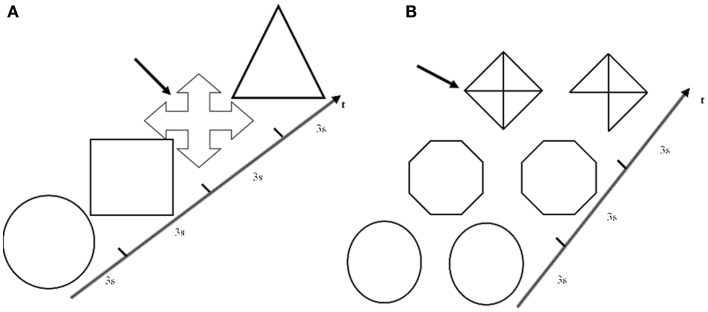
Eleven right-handed non-demented right-sided onset PD patients without visuospatial impairment or hallucinations and 11 healthy controls were studied with functional magnetic resonance imaging while performing a specific visuoperceptual/visuospatial paradigm that allowed to highlight the specific process underlying visuospatial judgment.
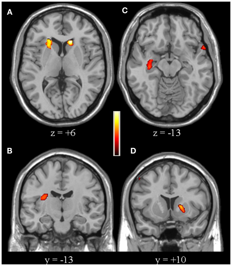
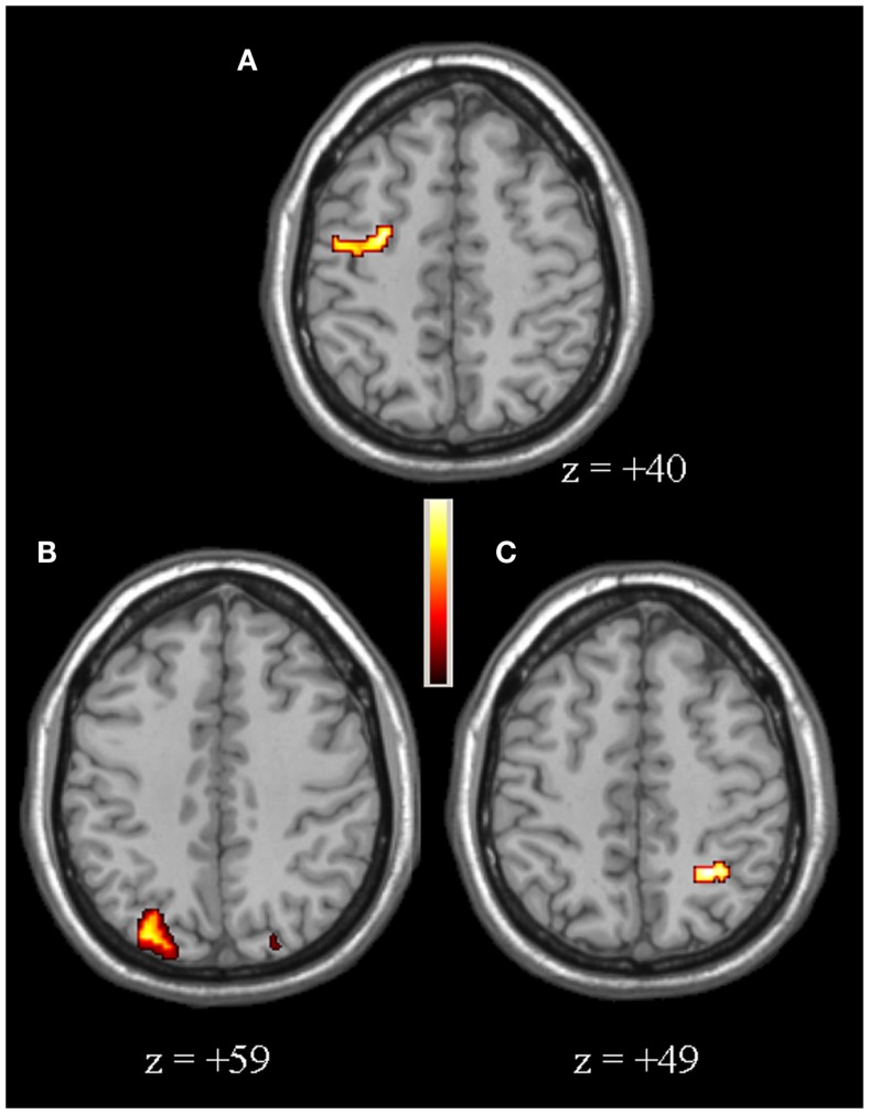
Significant changes in both cortical areas and subcortical regions involved in visual stimuli processing were observed. In particular, PD patients showed a reduced activation for the right insula, left putamen, bilateral caudate, and right hippocampus, as well as an over-activation of the right dorso-lateral prefrontal and of the posterior parietal cortices, particularly in the right hemisphere.
We found that both loss of efficiency and compensatory mechanisms occur in PD patients, providing further insight into the pathophysiological role of the functional alterations of basal ganglia and limbic structures in the impairment of visuoperceptual and visuospatial functions observed in PD.
视觉感知缺陷是帕金森病(PD)的常见表现。最近,在伴有认知衰退和视幻觉的PD患者中观察到,与视觉刺激分析相关的额顶叶区域和皮质下区域存在结构异常。本研究的目的是调查认知未受损的PD患者视觉感知的显著特征。
对11名无视觉空间障碍或幻觉的右利手、右侧起病的非痴呆PD患者和11名健康对照者进行功能磁共振成像研究,同时让他们执行一种特定的视觉感知/视觉空间范式,以突出视觉空间判断的具体过程。
观察到参与视觉刺激处理的皮质区域和皮质下区域均有显著变化。具体而言,PD患者右侧岛叶、左侧壳核、双侧尾状核和右侧海马的激活减少,右侧背外侧前额叶和顶叶后皮质过度激活,尤其是在右半球。
我们发现PD患者存在效率丧失和代偿机制,这为深入了解基底神经节和边缘结构功能改变在PD患者视觉感知和视觉空间功能损害中的病理生理作用提供了进一步的见解。