Zhang Ye, Zhang Lianyang, Li Yang, Sun Shijin, Tan Hao
State Key Laboratory of Trauma, Burns and Combined Injury, Institute of Surgery Research, Daping Hospital, Third Military Medical University, Chongqing, China.
PLoS One. 2014 Sep 2;9(9):e106328. doi: 10.1371/journal.pone.0106328. eCollection 2014.
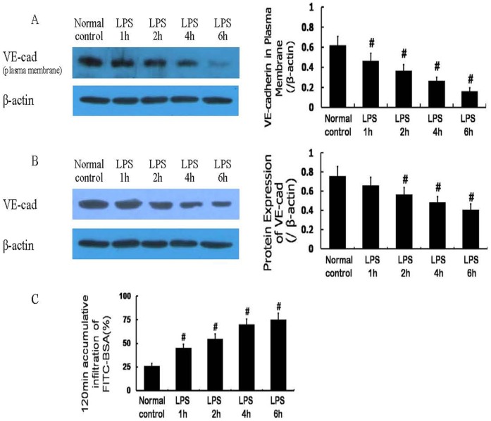
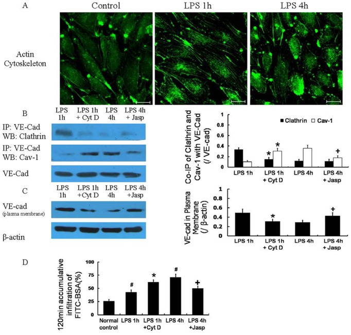
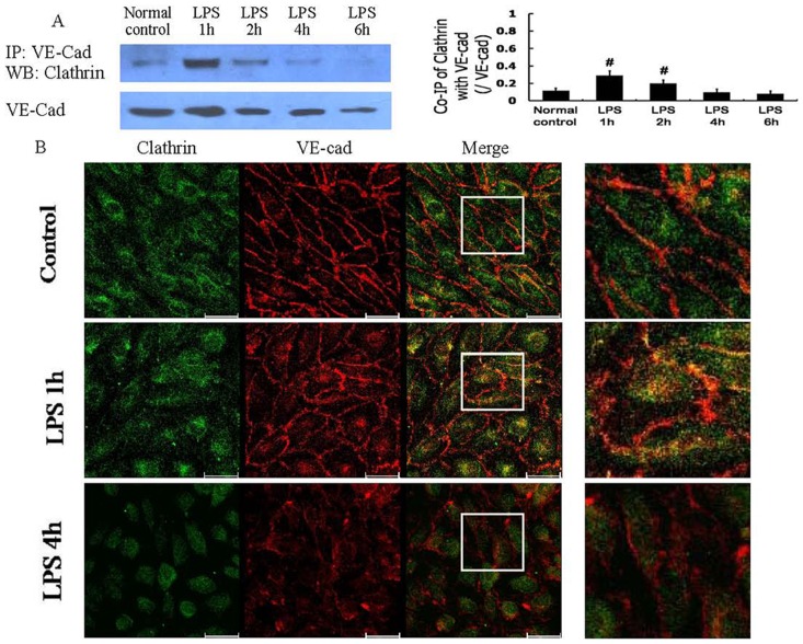
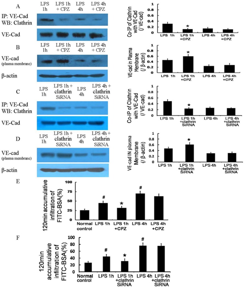
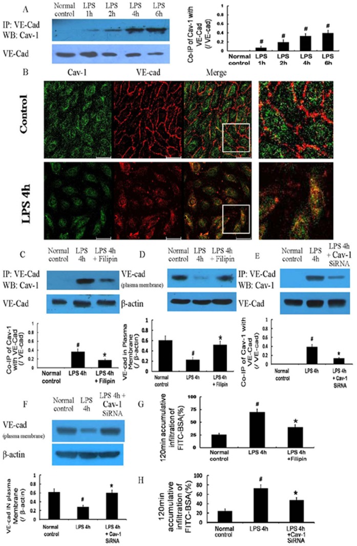
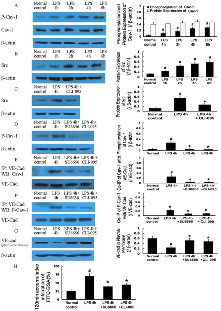
Vascular hyperpermeability induced by lipopolysaccharide (LPS) is a common pathogenic process in cases of severe trauma and sepsis. Vascular endothelial cadherin (VE-cad) is a key regulatory molecule involved in this process, although the detailed mechanism through which this molecule acts remains unclear. We assessed the role of clathrin-mediated and caveolae-mediated endocytosis of VE-cad in LPS-induced vascular hyperpermeability in the human vascular endothelial cell line CRL-2922 and determined that vascular permeability and VE-cad localization at the plasma membrane were negatively correlated after LPS treatment. Additionally, the loss of VE-cad at the plasma membrane was caused by both clathrin-mediated and caveolae-mediated endocytosis. Clathrin-mediated endocytosis was dominant early after LPS treatment, and caveolae-mediated endocytosis was dominant hours after LPS treatment. The caveolae-mediated endocytosis of VE-cad was activated through the LPS-Toll-like receptor 4 (TLR4)-Src signaling pathway. Structural changes in the actin cytoskeleton, specifically from polymerization to depolymerization, were important reasons for the switching of the VE-cad endocytosis pathway from clathrin-mediated to caveolae-mediated. Our findings suggest that clathrin-mediated and caveolae-mediated endocytosis of VE-cad contribute to LPS-induced vascular hyperpermeability, although they contribute via different mechanism. The predominant means of endocytosis depends on the time since LPS treatment.
脂多糖(LPS)诱导的血管通透性增加是严重创伤和脓毒症病例中常见的致病过程。血管内皮钙黏蛋白(VE-cad)是参与该过程的关键调节分子,但其作用的详细机制尚不清楚。我们评估了网格蛋白介导的和小窝介导的VE-cad内吞作用在人血管内皮细胞系CRL-2922中LPS诱导的血管通透性增加中的作用,并确定LPS处理后血管通透性与质膜上VE-cad的定位呈负相关。此外,质膜上VE-cad的丢失是由网格蛋白介导的和小窝介导的内吞作用共同导致的。LPS处理后早期,网格蛋白介导的内吞作用占主导,而LPS处理数小时后,小窝介导的内吞作用占主导。VE-cad的小窝介导的内吞作用通过LPS- Toll样受体4(TLR4)-Src信号通路被激活。肌动蛋白细胞骨架的结构变化,特别是从聚合到解聚,是VE-cad内吞途径从网格蛋白介导转变为小窝介导的重要原因。我们的研究结果表明,VE-cad的网格蛋白介导的和小窝介导的内吞作用均有助于LPS诱导的血管通透性增加,尽管它们通过不同的机制起作用。内吞作用的主要方式取决于LPS处理后的时间。