D'Souza Dany V, Jonckers Elisabeth, Bruns Andreas, Künnecke Basil, von Kienlin Markus, Van der Linden Annemie, Mueggler Thomas, Verhoye Marleen
F. Hoffmann-La Roche Pharmaceuticals Ltd, Neuroscience Discovery, Basel, Switzerland.
Bio-Imaging Lab, University of Antwerp, Antwerp, Belgium.
PLoS One. 2014 Sep 2;9(9):e106156. doi: 10.1371/journal.pone.0106156. eCollection 2014.
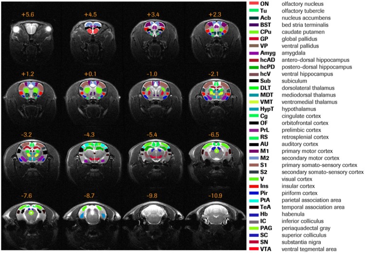
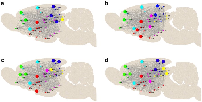
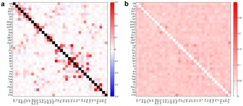
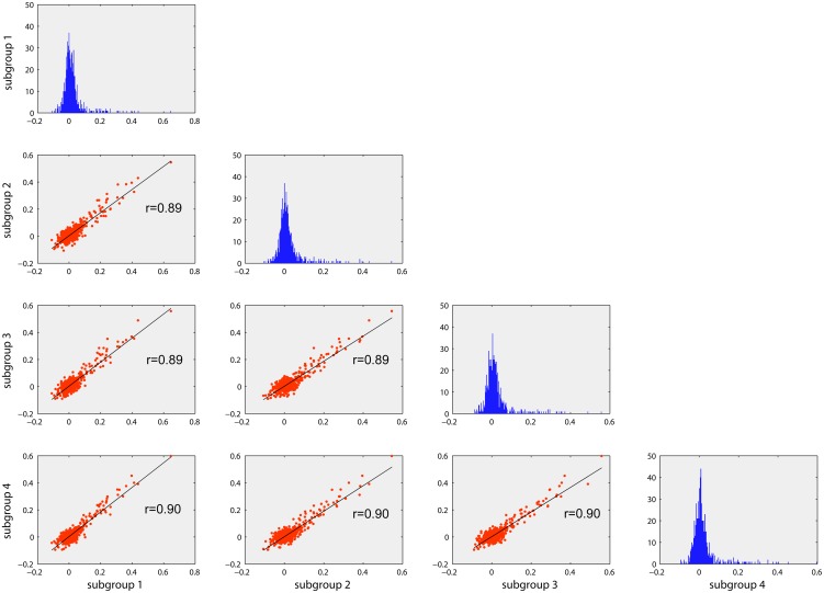
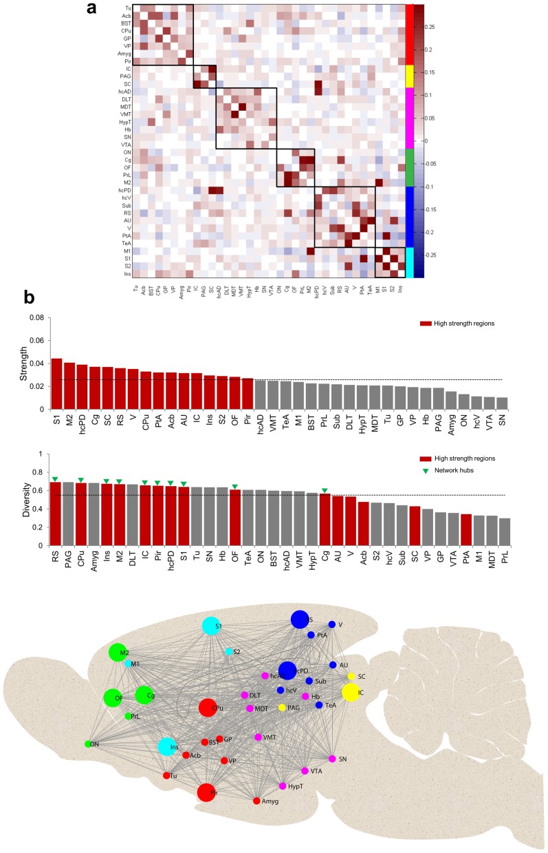
Translation of resting-state functional connectivity (FC) magnetic resonance imaging (rs-fMRI) applications from human to rodents has experienced growing interest, and bears a great potential in pre-clinical imaging as it enables assessing non-invasively the topological organization of complex FC networks (FCNs) in rodent models under normal and various pathophysiological conditions. However, to date, little is known about the organizational architecture of FCNs in rodents in a mentally healthy state, although an understanding of the same is of paramount importance before investigating networks under compromised states. In this study, we characterized the properties of resting-state FCN in an extensive number of Sprague-Dawley rats (n = 40) under medetomidine sedation by evaluating its modular organization and centrality of brain regions and tested for reproducibility. Fully-connected large-scale complex networks of positively and negatively weighted connections were constructed based on Pearson partial correlation analysis between the time courses of 36 brain regions encompassing almost the entire brain. Applying recently proposed complex network analysis measures, we show that the rat FCN exhibits a modular architecture, comprising six modules with a high between subject reproducibility. In addition, we identified network hubs with strong connections to diverse brain regions. Overall our results obtained under a straight medetomidine protocol show for the first time that the community structure of the rat brain is preserved under pharmacologically induced sedation with a network modularity contrasting from the one reported for deep anesthesia but closely resembles the organization described for the rat in conscious state.
从人类到啮齿动物的静息态功能连接(FC)磁共振成像(rs-fMRI)应用的翻译受到越来越多的关注,并且在临床前成像中具有巨大潜力,因为它能够在正常和各种病理生理条件下非侵入性地评估啮齿动物模型中复杂FC网络(FCN)的拓扑组织。然而,迄今为止,对于处于精神健康状态的啮齿动物中FCN的组织结构知之甚少,尽管在研究受损状态下的网络之前了解这一点至关重要。在本研究中,我们通过评估其模块化组织和脑区中心性,对大量在美托咪定镇静下的Sprague-Dawley大鼠(n = 40)的静息态FCN特性进行了表征,并测试了其可重复性。基于涵盖几乎整个大脑的36个脑区时间序列之间的Pearson偏相关分析,构建了具有正加权和负加权连接的全连接大规模复杂网络。应用最近提出的复杂网络分析方法,我们表明大鼠FCN呈现出模块化结构,由六个模块组成,个体间具有高度可重复性。此外,我们识别出与不同脑区有强连接的网络枢纽。总体而言,我们在直接美托咪定方案下获得的结果首次表明,在药物诱导的镇静下大鼠脑的群落结构得以保留,其网络模块化与深度麻醉下报道的不同,但与清醒状态下大鼠的组织描述非常相似。