• 文献检索
  • 文档翻译
  • 深度研究
  • 学术资讯
  • Suppr Zotero 插件Zotero 插件
  • 邀请有礼
  • 套餐&价格
  • 历史记录
应用&插件
Suppr Zotero 插件Zotero 插件浏览器插件Mac 客户端Windows 客户端微信小程序
定价
高级版会员购买积分包购买API积分包
服务
文献检索文档翻译深度研究API 文档MCP 服务
关于我们
关于 Suppr公司介绍联系我们用户协议隐私条款
关注我们

Suppr 超能文献

核心技术专利:CN118964589B侵权必究
粤ICP备2023148730 号-1Suppr @ 2026

文献检索

告别复杂PubMed语法,用中文像聊天一样搜索,搜遍4000万医学文献。AI智能推荐,让科研检索更轻松。

立即免费搜索

文件翻译

保留排版,准确专业,支持PDF/Word/PPT等文件格式,支持 12+语言互译。

免费翻译文档

深度研究

AI帮你快速写综述,25分钟生成高质量综述,智能提取关键信息,辅助科研写作。

立即免费体验

血管生成和造血细胞祖细胞分散在产前小鼠心脏内。

Vasculogenic and hematopoietic cellular progenitors are scattered within the prenatal mouse heart.

作者信息

Jankowska-Steifer Ewa, Madej Maria, Niderla-Bielińska Justyna, Ruminski Sławomir, Flaht-Zabost Aleksandra, Czarnowska Elzbieta, Gula Grzegorz, Radomska-Leśniewska Dorota M, Ratajska Anna

机构信息

Department of Histology and Embryology, Medical University of Warsaw, Warsaw, Poland,

出版信息

Histochem Cell Biol. 2015 Feb;143(2):153-69. doi: 10.1007/s00418-014-1269-z. Epub 2014 Sep 9.

DOI:10.1007/s00418-014-1269-z
PMID:25201347
原文链接:https://pmc.ncbi.nlm.nih.gov/articles/PMC4298664/
Abstract

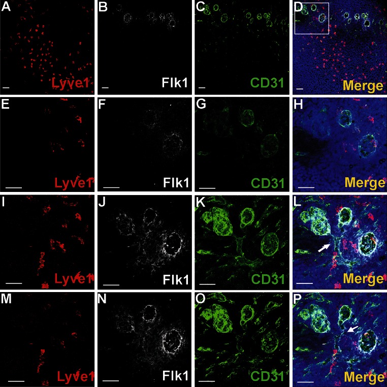
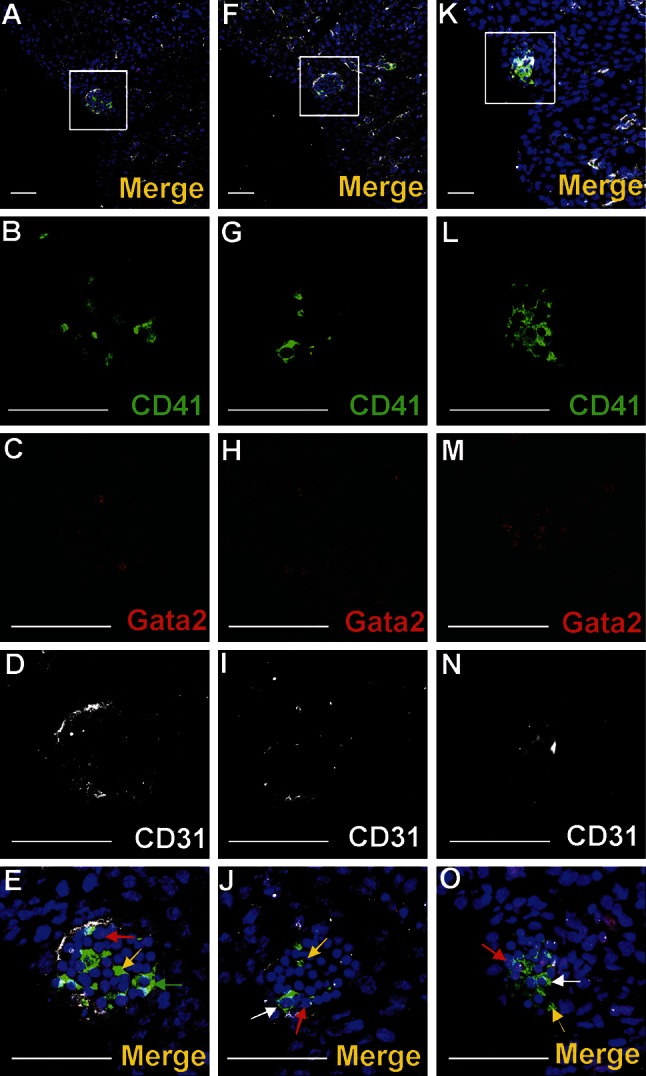
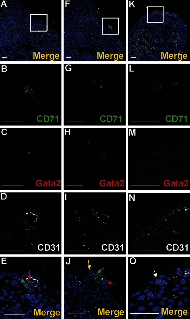
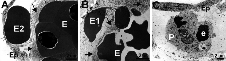
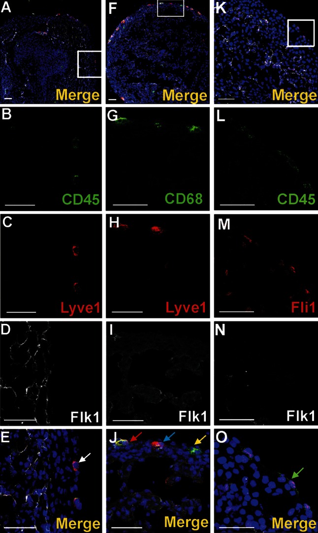
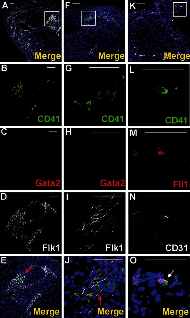
Vasculogenesis and hematopoiesis are co-localized in the embryonic body, but precise phenotypes of the cells contributing to these processes are not defined. The aim of this study was to characterize phenotypic profiles and location of putative vasculogenic and hematopoietic cellular progenitors in the embryonic mouse heart. Confocal microscopy, as well as ultrastructural and stereomicroscopic analyses, was performed on immunohistochemical whole-mount-stained or sectioned hearts at stages 11.5-14 dpc. A FASC analysis was conducted to quantify putative vasculogenic and hematopoietic cells. We found subepicardial blood islands in the form of foci of accumulation of cells belonging to erythroblastic and megakaryocytic lineages at various stages of maturation, exhibiting phenotypes: GATA2(+)/CD41(+), GATA2(-)/CD41(+), GATA2(+)/CD71(-), GATA2(-)/CD71(+), Fli1(+)/CD71(+), Fli1(-)/CD71(+), with a majority of cells expressing the Ter119 antigen, but none of them expressing Flk1. The subepicardium and the outflow tract endothelium were recognized to be the areas where progenitor cells were scattered or adjoining the endothelial cells. These progenitor cells were characterized as possessing the following antigens: CD45(+)/Fli1(+), CD41(+)/Flk1(+), Flk1(+)/Fli1(+). A FACS analysis demonstrated that the CD41/Flk1 double-positive population of cells constituted 2.68% of total cell population isolated from 12.5 dpc hearts. Vessels and tubules were positive for CD31, Flk1, Fli1, Tie2, including blood islands endothelia. The endocardial wall endothelia were found to function as an anchoring apparatus for megakaryocytes releasing platelets into the cardiac cavities. Phenotypic characteristics of vasculogenic (Flk1(+)/Fli1(+)) and hematopoietic (GATA2(+)/CD71(+), CD41(+)/GATA2(+)) progenitors, as well as the putative hemogenic endothelium (Flk1(+)/CD41(+)) in embryonic mouse hearts, have been presented. Cardiac blood islands, the subepicardium and endothelium of the outflow tract cushions have been defined as areas where these progenitor cells can be found.

摘要

血管生成和造血作用在胚胎体内共定位,但参与这些过程的细胞的精确表型尚未明确。本研究的目的是表征胚胎小鼠心脏中假定的血管生成和造血细胞祖细胞的表型特征及位置。对胚胎发育第11.5 - 14天的免疫组织化学全层染色或切片心脏进行共聚焦显微镜检查以及超微结构和立体显微镜分析。进行FASC分析以量化假定的血管生成和造血细胞。我们发现心外膜下血岛,其形式为处于不同成熟阶段的属于成红细胞和巨核细胞谱系的细胞聚集灶,表现出以下表型:GATA2(+)/CD41(+)、GATA2(-)/CD41(+)、GATA2(+)/CD71(-)、GATA2(-)/CD71(+)、Fli1(+)/CD71(+)、Fli1(-)/CD71(+),大多数细胞表达Ter119抗原,但均不表达Flk1。心外膜下层和流出道内皮被认为是祖细胞散布或与内皮细胞相邻的区域。这些祖细胞的特征是具有以下抗原:CD45(+)/Fli1(+)、CD41(+)/Flk1(+)、Flk1(+)/Fli1(+)。FACS分析表明,CD41/Flk1双阳性细胞群体占从胚胎发育第12.5天心脏分离的总细胞群体的2.68%。血管和小管对CD31、Flk1、Fli1、Tie2呈阳性,包括血岛内的内皮。发现心内膜壁内皮作为巨核细胞向心腔释放血小板的锚定装置。本文呈现了胚胎小鼠心脏中血管生成祖细胞(Flk1(+)/Fli1(+))和造血祖细胞(GATA2(+)/CD71(+)、CD41(+)/GATA2(+))以及假定的造血内皮(Flk1(+)/CD41(+))的表型特征。心脏血岛、心外膜下层和流出道垫的内皮已被定义为可发现这些祖细胞的区域。

https://cdn.ncbi.nlm.nih.gov/pmc/blobs/9d77/4298664/f0afb2af7814/418_2014_1269_Fig15_HTML.jpg
https://cdn.ncbi.nlm.nih.gov/pmc/blobs/9d77/4298664/8048fe3f7ae0/418_2014_1269_Fig1_HTML.jpg
https://cdn.ncbi.nlm.nih.gov/pmc/blobs/9d77/4298664/a276f413a25f/418_2014_1269_Fig2_HTML.jpg
https://cdn.ncbi.nlm.nih.gov/pmc/blobs/9d77/4298664/68e4f7fe5e96/418_2014_1269_Fig3_HTML.jpg
https://cdn.ncbi.nlm.nih.gov/pmc/blobs/9d77/4298664/171e189968de/418_2014_1269_Fig4_HTML.jpg
https://cdn.ncbi.nlm.nih.gov/pmc/blobs/9d77/4298664/03f059f65a25/418_2014_1269_Fig5_HTML.jpg
https://cdn.ncbi.nlm.nih.gov/pmc/blobs/9d77/4298664/6ffd91419860/418_2014_1269_Fig6_HTML.jpg
https://cdn.ncbi.nlm.nih.gov/pmc/blobs/9d77/4298664/77dbe4d769a1/418_2014_1269_Fig7_HTML.jpg
https://cdn.ncbi.nlm.nih.gov/pmc/blobs/9d77/4298664/7c0248810aff/418_2014_1269_Fig8_HTML.jpg
https://cdn.ncbi.nlm.nih.gov/pmc/blobs/9d77/4298664/902e3029925e/418_2014_1269_Fig9_HTML.jpg
https://cdn.ncbi.nlm.nih.gov/pmc/blobs/9d77/4298664/d1aca511dc70/418_2014_1269_Fig10_HTML.jpg
https://cdn.ncbi.nlm.nih.gov/pmc/blobs/9d77/4298664/995b85314d79/418_2014_1269_Fig11_HTML.jpg
https://cdn.ncbi.nlm.nih.gov/pmc/blobs/9d77/4298664/c9c9bfc49cce/418_2014_1269_Fig12_HTML.jpg
https://cdn.ncbi.nlm.nih.gov/pmc/blobs/9d77/4298664/4bbd92b01664/418_2014_1269_Fig13_HTML.jpg
https://cdn.ncbi.nlm.nih.gov/pmc/blobs/9d77/4298664/2acd4379d082/418_2014_1269_Fig14_HTML.jpg
https://cdn.ncbi.nlm.nih.gov/pmc/blobs/9d77/4298664/f0afb2af7814/418_2014_1269_Fig15_HTML.jpg

相似文献

1
Vasculogenic and hematopoietic cellular progenitors are scattered within the prenatal mouse heart.血管生成和造血细胞祖细胞分散在产前小鼠心脏内。
Histochem Cell Biol. 2015 Feb;143(2):153-69. doi: 10.1007/s00418-014-1269-z. Epub 2014 Sep 9.
2
Spry1 is expressed in hemangioblasts and negatively regulates primitive hematopoiesis and endothelial cell function.Spry1 在血岛细胞中表达,并负调控原始造血和内皮细胞功能。
PLoS One. 2011 Apr 1;6(4):e18374. doi: 10.1371/journal.pone.0018374.
3
Endothelial cells in the early murine yolk sac give rise to CD41-expressing hematopoietic cells.早期小鼠卵黄囊中的内皮细胞可产生表达CD41的造血细胞。
Stem Cells Dev. 2005 Feb;14(1):44-54. doi: 10.1089/scd.2005.14.44.
4
Cells with hematopoietic potential reside within mouse proepicardium.具有造血潜能的细胞存在于小鼠的心外膜前体细胞中。
Histochem Cell Biol. 2018 Jun;149(6):577-591. doi: 10.1007/s00418-018-1661-1. Epub 2018 Mar 16.
5
Progressive lineage analysis by cell sorting and culture identifies FLK1+VE-cadherin+ cells at a diverging point of endothelial and hemopoietic lineages.通过细胞分选和培养进行的渐进谱系分析在内皮细胞谱系和造血细胞谱系的分歧点鉴定出了FLK1+VE-钙黏蛋白+细胞。
Development. 1998 May;125(9):1747-57. doi: 10.1242/dev.125.9.1747.
6
CD45 expression discriminates waves of embryonic megakaryocytes in the mouse.CD45 表达可区分小鼠胚胎巨核细胞的波。
Haematologica. 2019 Sep;104(9):1853-1865. doi: 10.3324/haematol.2018.192559. Epub 2018 Dec 20.
7
Expression of CD41 marks the initiation of definitive hematopoiesis in the mouse embryo.CD41的表达标志着小鼠胚胎中确定性造血的起始。
Blood. 2003 Jan 15;101(2):508-16. doi: 10.1182/blood-2002-06-1699. Epub 2002 Sep 19.
8
Distinct hemogenic potential of endothelial cells and CD41+ cells in mouse embryos.小鼠胚胎中内皮细胞和CD41+细胞不同的造血潜能。
Dev Growth Differ. 2007 May;49(4):287-300. doi: 10.1111/j.1440-169X.2007.00925.x.
9
The heart endocardium is derived from vascular endothelial progenitors.心脏的心内膜来源于血管内皮祖细胞。
Development. 2011 Nov;138(21):4777-87. doi: 10.1242/dev.061192.
10
[Study on Flk1 Cells during Mouse Early Embryogenesis by Lineage Tracing].通过谱系追踪对小鼠早期胚胎发育过程中Flk1细胞的研究
Zhongguo Shi Yan Xue Ye Xue Za Zhi. 2019 Jun;27(3):942-949. doi: 10.19746/j.cnki.issn.1009-2137.2019.03.050.

引用本文的文献

1
Notch and retinoic acid signals regulate macrophage formation from endocardium downstream of Nkx2-5.Notch 和视黄酸信号调节 Nkx2-5 下游的心内膜向巨噬细胞的形成。
Nat Commun. 2023 Sep 5;14(1):5398. doi: 10.1038/s41467-023-41039-6.
2
Sox7-positive endothelial progenitors establish coronary arteries and govern ventricular compaction.Sox7 阳性的内皮祖细胞建立冠状动脉,并控制心室的致密化。
EMBO Rep. 2023 Oct 9;24(10):e55043. doi: 10.15252/embr.202255043. Epub 2023 Aug 8.
3
Desmoglein 2 regulates cardiogenesis by restricting hematopoiesis in the developing murine heart.

本文引用的文献

1
Induction of the Proepicardium.心外膜原基的诱导
J Dev Biol. 2013 Sep 1;1(2):82-91. doi: 10.3390/jdb1020082.
2
Macrophage biology in development, homeostasis and disease.发育、稳态和疾病中的巨噬细胞生物学。
Nature. 2013 Apr 25;496(7446):445-55. doi: 10.1038/nature12034.
3
Haemogenic endocardium contributes to transient definitive haematopoiesis.血发生心内膜有助于短暂的终末性造血。
桥粒芯糖蛋白 2 通过限制发育中鼠心脏的造血作用来调节心脏发生。
Sci Rep. 2021 Nov 4;11(1):21687. doi: 10.1038/s41598-021-00996-y.
4
Monomeric C-reactive protein via endothelial CD31 for neurovascular inflammation in an ApoE genotype-dependent pattern: A risk factor for Alzheimer's disease?单体 C 反应蛋白通过血管内皮细胞 CD31 介导的神经血管炎症的 ApoE 基因型依赖性模式:阿尔茨海默病的危险因素?
Aging Cell. 2021 Nov;20(11):e13501. doi: 10.1111/acel.13501. Epub 2021 Oct 23.
5
Potential functions of embryonic cardiac macrophages in angiogenesis, lymphangiogenesis and extracellular matrix remodeling.胚胎心脏巨噬细胞在血管生成、淋巴管生成和细胞外基质重塑中的潜在功能。
Histochem Cell Biol. 2021 Jan;155(1):117-132. doi: 10.1007/s00418-020-01934-1. Epub 2020 Nov 1.
6
Arterial identity of hemogenic endothelium: a key to unlock definitive hematopoietic commitment in human pluripotent stem cell cultures.造血内皮细胞的动脉特性:开启人类多能干细胞培养中确定造血定向分化的关键。
Exp Hematol. 2019 Mar;71:3-12. doi: 10.1016/j.exphem.2018.11.007. Epub 2018 Nov 28.
7
Cells with hematopoietic potential reside within mouse proepicardium.具有造血潜能的细胞存在于小鼠的心外膜前体细胞中。
Histochem Cell Biol. 2018 Jun;149(6):577-591. doi: 10.1007/s00418-018-1661-1. Epub 2018 Mar 16.
8
Insights into blood cell formation from hemogenic endothelium in lesser-known anatomic sites.对鲜为人知的解剖部位中造血内皮细胞形成血细胞的见解。
Dev Dyn. 2016 Oct;245(10):1011-28. doi: 10.1002/dvdy.24430. Epub 2016 Aug 17.
9
Extravascular endothelial and hematopoietic islands form through multiple pathways in midgestation mouse embryos.血管外内皮细胞和造血岛在妊娠中期小鼠胚胎中通过多种途径形成。
Dev Biol. 2016 Jul 1;415(1):111-121. doi: 10.1016/j.ydbio.2016.04.004. Epub 2016 Apr 20.
10
The Histochemistry and Cell Biology omnium-gatherum: the year 2015 in review.《组织化学与细胞生物学综述:2015年回顾》
Histochem Cell Biol. 2016 Mar;145(3):239-74. doi: 10.1007/s00418-016-1417-8. Epub 2016 Feb 15.
Nat Commun. 2013;4:1564. doi: 10.1038/ncomms2569.
4
Hemogenic endothelium: a vessel for blood production.血发生内皮:造血的血管。
Int J Biochem Cell Biol. 2013 Mar;45(3):692-5. doi: 10.1016/j.biocel.2012.12.013. Epub 2012 Dec 25.
5
Mouse embryonic head as a site for hematopoietic stem cell development.小鼠胚胎头部作为造血干细胞发育的部位。
Cell Stem Cell. 2012 Nov 2;11(5):663-75. doi: 10.1016/j.stem.2012.07.004.
6
Cellular phenotypes and spatio-temporal patterns of lymphatic vessel development in embryonic mouse hearts.胚胎鼠心脏淋巴管发育的细胞表型和时空模式。
Dev Dyn. 2012 Sep;241(9):1473-86. doi: 10.1002/dvdy.23827. Epub 2012 Jul 19.
7
Hemogenic endothelium during development and beyond.发育中和发育后的造血内皮。
Blood. 2012 May 24;119(21):4823-7. doi: 10.1182/blood-2011-12-353466. Epub 2012 Mar 13.
8
Whole-mount confocal microscopy for vascular branching morphogenesis.用于血管分支形态发生的整体共聚焦显微镜检查。
Methods Mol Biol. 2012;843:69-78. doi: 10.1007/978-1-61779-523-7_7.
9
Release of angiogenesis regulatory proteins from platelet alpha granules: modulation of physiologic and pathologic angiogenesis.血小板α 颗粒中血管生成调节蛋白的释放:对生理性和病理性血管生成的调节。
Blood. 2011 Aug 4;118(5):1359-69. doi: 10.1182/blood-2011-02-334524. Epub 2011 Jun 16.
10
Coronary arteries form by developmental reprogramming of venous cells.冠状动脉通过静脉细胞的发育重编程形成。
Nature. 2010 Mar 25;464(7288):549-53. doi: 10.1038/nature08873.