Nguyen-Ngoc Kim-Vy, Shamir Eliah R, Huebner Robert J, Beck Jennifer N, Cheung Kevin J, Ewald Andrew J
Departments of Cell Biology and Oncology, Center for Cell Dynamics, Johns Hopkins School of Medicine, 855 N. Wolfe Street, 452 Rangos Building, Baltimore, MD, 21205, USA.
Methods Mol Biol. 2015;1189:135-62. doi: 10.1007/978-1-4939-1164-6_10.
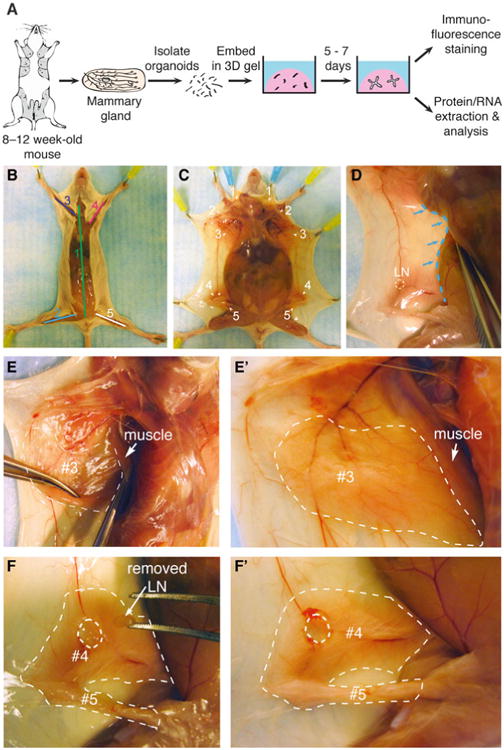
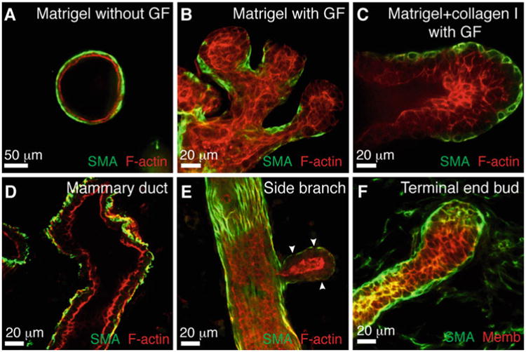
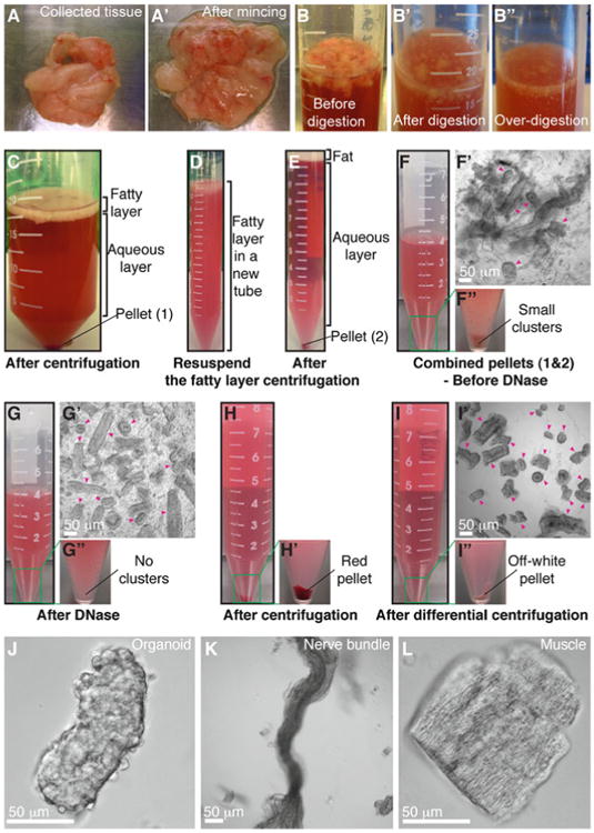
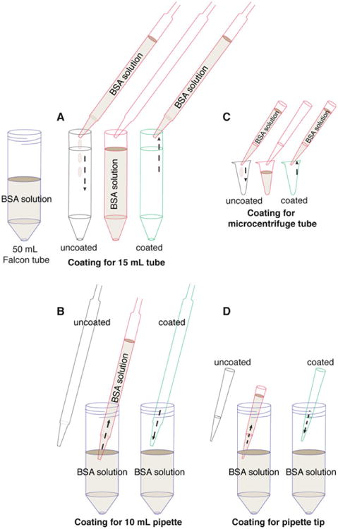
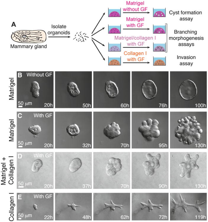
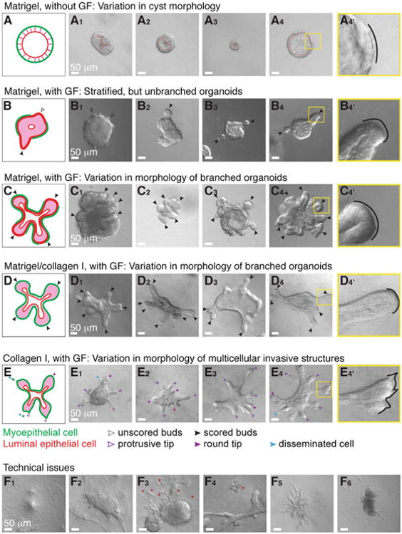
Epithelia are fundamental tissues that line cavities, glands, and outer body surfaces. We use three-dimensional (3D) embedded culture of primary murine mammary epithelial ducts, called "organoids," to recapitulate in days in culture epithelial programs that occur over weeks deep within the body. Modulating the composition of the extracellular matrix (ECM) allows us to model cell- and tissue-level behaviors observed in normal development, such as branching morphogenesis, and in cancer, such as invasion and dissemination. Here, we describe a collection of protocols for 3D culture of mammary organoids in different ECMs and for immunofluorescence staining of 3D culture samples and mammary gland tissue sections. We illustrate expected phenotypic outcomes of each assay and provide troubleshooting tips for commonly encountered technical problems.
上皮组织是覆盖在腔、腺体和身体外表面的基本组织。我们使用原代小鼠乳腺上皮导管的三维(3D)包埋培养,即所谓的“类器官”,在数天的培养中重现那些在体内深处数周才会发生的上皮程序。调节细胞外基质(ECM)的组成使我们能够模拟在正常发育中观察到的细胞和组织水平行为,如分支形态发生,以及在癌症中观察到的行为,如侵袭和扩散。在这里,我们描述了一系列用于在不同ECM中进行乳腺类器官3D培养以及对3D培养样品和乳腺组织切片进行免疫荧光染色的方案。我们展示了每种检测预期的表型结果,并针对常见的技术问题提供了故障排除提示。