Akbari Sari Ali, Mobinizadeh Mohammadreza, Azadbakht Mahdi
Dept. of Health Management and Economics, Tehran University of Medical Sciences, Tehran, Iran.
Dept. of Health Service Management, Science and Research Branch, Islamic Azad University, Tehran, Iran.
Iran J Cancer Prev. 2013 Winter;6(1):44-51.
Optical mammography is a new diagnostic method that uses Near-infrared for detection of functional abnormalities and shows tissue activities by measuring absorption and scattering of Near-infrared light. This study aims to evaluate the safety and effectiveness of this technology.
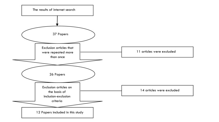
Cochrane Library (Issue 10, 2012) and Medline (Nov 2012) weresearched using free text and Mesh. Studies that compared optical mammography with other diagnostic methods and used outcomes such as sensitivity, specificity and safety were included.
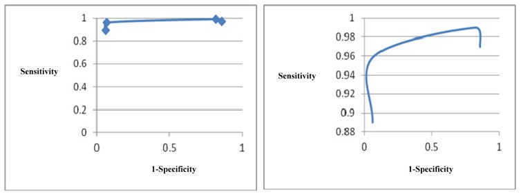
Twelve studies were included in this review. A multicenter RCT showed that among 875 biopsied lesions, suspicion index led to 97% sensitivity, 14%specificity, 95% negative predictive value and 24% positive predictive value. In terms of oxygenation index, the included studies found that the process should be used with various wavelengths compared to single wavelength technique (690, 750, 788, 856 nm or 683, 912, 975nm). In terms of sensitivity and specificity, Diffuse Optical Tomography Computer Aided Detection is capable of distinguishing healthy tissues from malignant ones with 89% sensitivity and 94% specificity. Also, this technology could show increased blood flow around the tumor tissue compared to the healthy tissue effectively. Included studies did not report any information about the effects of technology on changing the treatment process or the final health outcomes.
Optical mammography is a safe, noninvasive, non-ionized diagnostic technology that can be used as a diagnostic supplement alongside conventional mammography for differentiating benign and malignant tumors. Women with higher breast density should be screened at younger ages and with more persistence than those who have lower densities.
光学乳腺造影是一种新的诊断方法,它利用近红外光检测功能异常,并通过测量近红外光的吸收和散射来显示组织活性。本研究旨在评估该技术的安全性和有效性。
使用自由文本和医学主题词(Mesh)检索Cochrane图书馆(2012年第10期)和Medline(2012年11月)。纳入比较光学乳腺造影与其他诊断方法并使用敏感性、特异性和安全性等结果的研究。
本综述纳入了12项研究。一项多中心随机对照试验表明,在875个经活检的病变中,可疑指数导致97%的敏感性、14%的特异性、95%的阴性预测值和24%的阳性预测值。就氧合指数而言,纳入的研究发现,与单波长技术(690、750、788、856纳米或683、912、975纳米)相比,该过程应使用各种波长。就敏感性和特异性而言,扩散光学断层扫描计算机辅助检测能够以89%的敏感性和94%的特异性区分健康组织和恶性组织。此外,与健康组织相比,该技术能有效显示肿瘤组织周围血流增加。纳入的研究未报告该技术对改变治疗过程或最终健康结局影响的任何信息。
光学乳腺造影是一种安全、无创、非电离的诊断技术,可作为传统乳腺造影的诊断补充,用于鉴别良性和恶性肿瘤。乳腺密度较高的女性应比密度较低的女性在更年轻时且更持续地进行筛查。