Nie Binbin, Liu Hua, Chen Kewei, Jiang Xiaofeng, Shan Baoci
Key Laboratory of Nuclear Analysis Techniques, Beijing Engineering Research Center of Radiographic Techniques and Equipment, Institute of High Energy Physics, Chinese Academy of Sciences, Beijing, China.
Banner Alzheimer's Institute, Banner Good Samaritan Positron Emission Tomography Center, Phoenix, Arizona, United States of America; Department of Mathematics and Statistics, Arizona State University, Tempe, Arizona, United States of America; Department of Radiology, University of Arizona, Tucson, Arizona, United States of America; Arizona Alzheimer's Consortium, Phoenix, Arizona, United States of America.
PLoS One. 2014 Sep 26;9(9):e108295. doi: 10.1371/journal.pone.0108295. eCollection 2014.
PET (positron emission tomography) imaging researches of functional metabolism using fluorodeoxyglucose (18F-FDG) of animal brain are important in neuroscience studies. FDG-PET imaging studies are often performed on groups of rats, so it is desirable to establish an objective voxel-based statistical methodology for group data analysis.
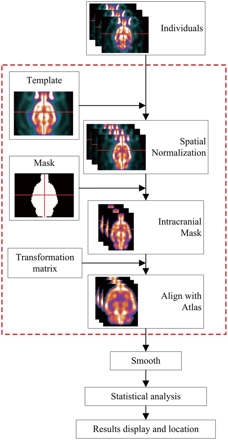
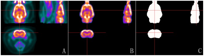
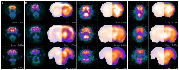
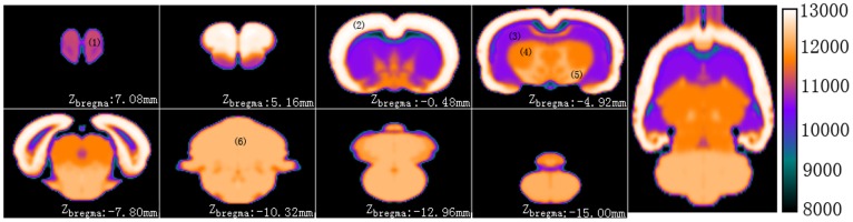
This study establishes a statistical parametric mapping (SPM) toolbox (plug-ins) named spmratIHEP for voxel-wise analysis of FDG-PET images of rat brain, in which an FDG-PET template and an intracranial mask image of rat brain in Paxinos & Watson space were constructed, and the default settings were modified according to features of rat brain. Compared to previous studies, our constructed rat brain template comprises not only the cerebrum and cerebellum, but also the whole olfactory bulb which made the later cognitive studies much more exhaustive. And with an intracranial mask image in the template space, the brain tissues of individuals could be extracted automatically. Moreover, an atlas space is used for anatomically labeling the functional findings in the Paxinos & Watson space. In order to standardize the template image with the atlas accurately, a synthetic FDG-PET image with six main anatomy structures is constructed from the atlas, which performs as a target image in the co-registration.
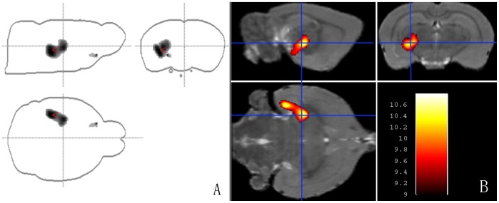
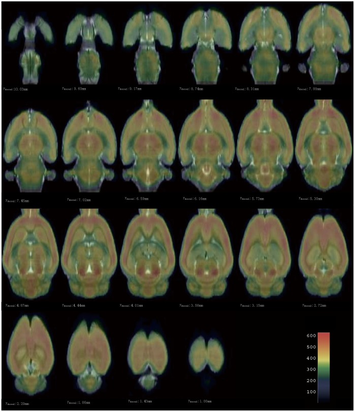
The spatial normalization procedure is evaluated, by which the individual rat brain images could be standardized into the Paxinos & Watson space successfully and the intracranial tissues could also be extracted accurately. The practical usability of this toolbox is evaluated using FDG-PET functional images from rats with left side middle cerebral artery occlusion (MCAO) in comparison to normal control rats. And the two-sample t-test statistical result is almost related to the left side MCA.
We established a toolbox of SPM8 named spmratIHEP for voxel-wise analysis of FDG-PET images of rat brain.
利用动物脑氟代脱氧葡萄糖(18F-FDG)进行功能代谢的正电子发射断层扫描(PET)成像研究在神经科学研究中具有重要意义。FDG-PET成像研究通常在大鼠群体上进行,因此需要建立一种基于体素的客观统计方法来进行群体数据分析。
本研究建立了一个名为spmratIHEP的统计参数映射(SPM)工具箱(插件),用于对大鼠脑FDG-PET图像进行逐体素分析,其中构建了Paxinos & Watson空间中大鼠脑的FDG-PET模板和颅内掩码图像,并根据大鼠脑的特征修改了默认设置。与以往研究相比,我们构建的大鼠脑模板不仅包括大脑和小脑,还包括整个嗅球,这使得后续的认知研究更加详尽。并且在模板空间中有颅内掩码图像,可以自动提取个体的脑组织。此外,使用图谱空间对Paxinos & Watson空间中的功能发现进行解剖学标记。为了使模板图像与图谱准确配准,从图谱构建了具有六个主要解剖结构的合成FDG-PET图像,该图像在配准中作为目标图像。
评估了空间归一化程序,通过该程序可以成功地将个体大鼠脑图像标准化到Paxinos & Watson空间,并且也可以准确提取颅内组织。使用左侧大脑中动脉闭塞(MCAO)大鼠与正常对照大鼠的FDG-PET功能图像评估了该工具箱的实际可用性。并且双样本t检验统计结果几乎与左侧MCA相关。
我们建立了一个名为spmratIHEP的SPM8工具箱,用于对大鼠脑FDG-PET图像进行逐体素分析。