Division of Nuclear Medicine, Department of Imaging and Pathology, KU Leuven, Leuven, Belgium.
PLoS One. 2013 Jun 7;8(6):e65286. doi: 10.1371/journal.pone.0065286. Print 2013.
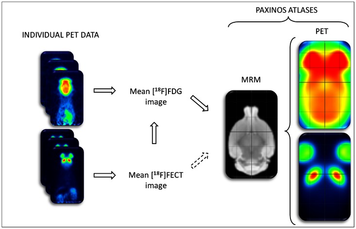
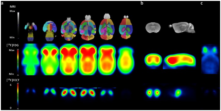
Automated voxel-based or pre-defined volume-of-interest (VOI) analysis of small-animal PET data in mice is necessary for optimal information usage as the number of available resolution elements is limited. We have mapped metabolic ([(18)F]FDG) and dopamine transporter ([(18)F]FECT) small-animal PET data onto a 3D Magnetic Resonance Microscopy (MRM) mouse brain template and aligned them in space to the Paxinos co-ordinate system. In this way, ligand-specific templates for sensitive analysis and accurate anatomical localization were created. Next, using a pre-defined VOI approach, test-retest and intersubject variability of various quantification methods were evaluated. Also, the feasibility of mouse brain statistical parametric mapping (SPM) was explored for [(18)F]FDG and [(18)F]FECT imaging of 6-hydroxydopamine-lesioned (6-OHDA) mice.
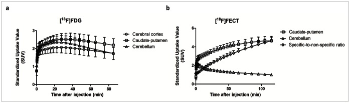
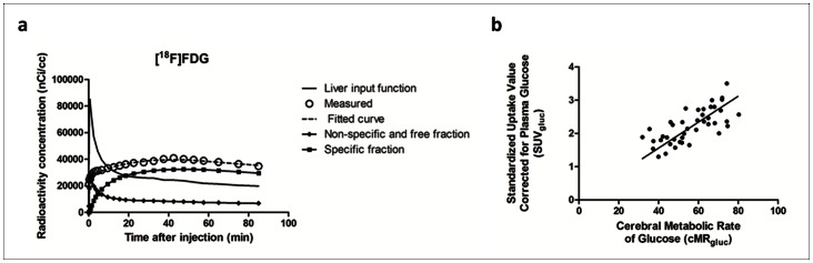
Twenty-three adult C57BL6 mice were scanned with [(18)F]FDG and [(18)F]FECT. Registrations and affine spatial normalizations were performed using SPM8. [(18)F]FDG data were quantified using (1) an image-derived-input function obtained from the liver (cMRglc), using (2) standardized uptake values (SUVglc) corrected for blood glucose levels and by (3) normalizing counts to the whole-brain uptake. Parametric [(18)F]FECT binding images were constructed by reference to the cerebellum. Registration accuracy was determined using random simulated misalignments and vectorial mismatch determination.
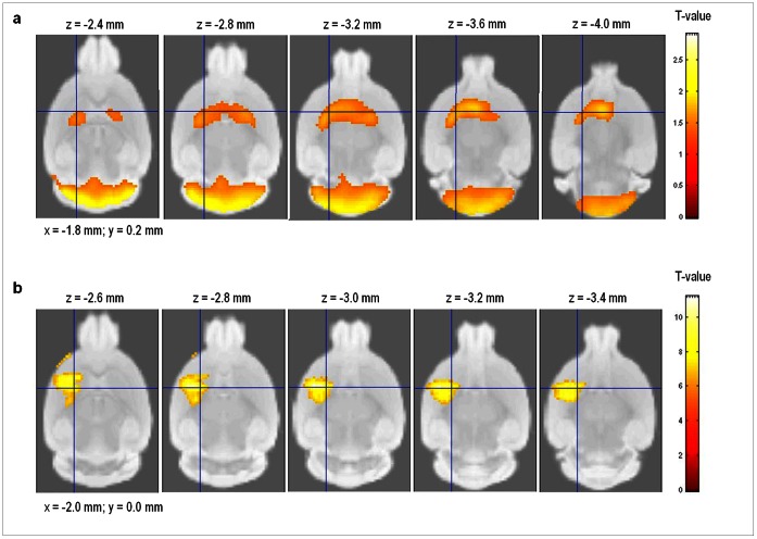
Registration accuracy was between 0.21-1.11 mm. Regional intersubject variabilities of cMRglc ranged from 15.4% to 19.2%, while test-retest values were between 5.0% and 13.0%. For [(18)F]FECT uptake in the caudate-putamen, these values were 13.0% and 10.3%, respectively. Regional values of cMRglc positively correlated to SUVglc measured within the 45-60 min time frame (spearman r = 0.71). Next, SPM analysis of 6-OHDA-lesioned mice showed hypometabolism in the bilateral caudate-putamen and cerebellum, and an unilateral striatal decrease in DAT availability.
MRM-based small-animal PET templates facilitate accurate assessment and spatial localization of mouse brain function using VOI or voxel-based analysis. Regional intersubject- and test-retest variations indicate that for these targets accuracy comparable to humans can be achieved.
为了最佳利用可用的分辨率元素数量有限的资源,在小动物 PET 数据中进行自动化体素或预定义的感兴趣区(VOI)分析对于获得最佳信息非常必要。我们已经将代谢([(18)F] FDG)和多巴胺转运蛋白([(18)F] FECT)的小动物 PET 数据映射到 3D 磁共振显微镜(MRM)小鼠脑模板上,并在空间上将它们与 Paxinos 坐标系统对齐。通过这种方式,创建了用于敏感分析和准确解剖定位的配体特异性模板。接下来,使用预定义的 VOI 方法,评估了各种定量方法的测试-重测和个体间变异性。此外,还探索了用于 6-羟多巴胺损伤(6-OHDA)小鼠的[(18)F] FDG 和[(18)F] FECT 成像的小鼠脑统计参数映射(SPM)的可行性。
对 23 只成年 C57BL6 小鼠进行了[(18)F] FDG 和[(18)F] FECT 扫描。使用 SPM8 进行配准和仿射空间归一化。[(18)F] FDG 数据使用以下方法进行定量:(1)从肝脏(cMRglc)获得的图像衍生输入函数,(2)校正血糖水平的标准化摄取值(SUVglc),以及(3)将计数归一化为全脑摄取。参考小脑构建了参数[(18)F] FECT 结合图像。使用随机模拟失配和矢量失配确定来确定配准精度。
配准精度在 0.21-1.11 毫米之间。cMRglc 的区域性个体间变异性范围为 15.4%至 19.2%,而测试-重测值范围为 5.0%至 13.0%。对于尾状核-壳核中的[(18)F] FECT 摄取,这些值分别为 13.0%和 10.3%。cMRglc 的区域值与在 45-60 分钟时间范围内测量的 SUVglc 呈正相关(Spearman r=0.71)。接下来,对 6-OHDA 损伤小鼠的 SPM 分析显示,双侧尾状核-壳核和小脑代谢降低,单侧纹状体 DAT 可用性降低。
基于 MRM 的小动物 PET 模板可通过 VOI 或体素分析促进对小鼠脑功能的准确评估和空间定位。区域性个体间和测试-重测变化表明,对于这些目标,可以达到与人类相当的准确性。