• 文献检索
  • 文档翻译
  • 深度研究
  • 学术资讯
  • Suppr Zotero 插件Zotero 插件
  • 邀请有礼
  • 套餐&价格
  • 历史记录
应用&插件
Suppr Zotero 插件Zotero 插件浏览器插件Mac 客户端Windows 客户端微信小程序
定价
高级版会员购买积分包购买API积分包
服务
文献检索文档翻译深度研究API 文档MCP 服务
关于我们
关于 Suppr公司介绍联系我们用户协议隐私条款
关注我们

Suppr 超能文献

核心技术专利:CN118964589B侵权必究
粤ICP备2023148730 号-1Suppr @ 2026

文献检索

告别复杂PubMed语法,用中文像聊天一样搜索,搜遍4000万医学文献。AI智能推荐,让科研检索更轻松。

立即免费搜索

文件翻译

保留排版,准确专业,支持PDF/Word/PPT等文件格式,支持 12+语言互译。

免费翻译文档

深度研究

AI帮你快速写综述,25分钟生成高质量综述,智能提取关键信息,辅助科研写作。

立即免费体验

肥厚型心肌病患者半自动延迟钆增强定量技术的准确性和可重复性

Accuracy and reproducibility of semi-automated late gadolinium enhancement quantification techniques in patients with hypertrophic cardiomyopathy.

作者信息

Mikami Yoko, Kolman Louis, Joncas Sebastien X, Stirrat John, Scholl David, Rajchl Martin, Lydell Carmen P, Weeks Sarah G, Howarth Andrew G, White James A

机构信息

Stephenson Cardiac Imaging Centre at the Libin Cardiovascular Institute of Alberta, University of Calgary, Calgary, AB, Canada.

Imaging Research Laboratory - Robarts Research Institute, Western University, London, ON, Canada.

出版信息

J Cardiovasc Magn Reson. 2014 Oct 7;16(1):85. doi: 10.1186/s12968-014-0085-x.

DOI:10.1186/s12968-014-0085-x
PMID:25315701
原文链接:https://pmc.ncbi.nlm.nih.gov/articles/PMC4189726/
Abstract

BACKGROUND

The presence and extent of late gadolinium enhancement (LGE) has been associated with adverse events in patients with hypertrophic cardiomyopathy (HCM). Signal intensity (SI) threshold techniques are routinely employed for quantification; Full-Width at Half-Maximum (FWHM) techniques are suggested to provide greater reproducibility than Signal Threshold versus Reference Mean (STRM) techniques, however the accuracy of these approaches versus the manual assignment of optimal SI thresholds has not been studied. In this study, we compared all known semi-automated LGE quantification techniques for accuracy and reproducibility among patients with HCM.

METHODS

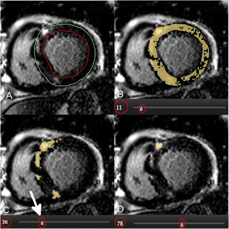
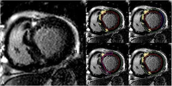
Seventy-six HCM patients (51 male, age 54 ± 13 years) were studied. Total LGE volume was quantified using 7 semi-automated techniques and compared to expert manual adjustment of the SI threshold to achieve optimal segmentation. Techniques tested included STRM based thresholds of >2, 3, 4, 5 and 6 SD above mean SI of reference myocardium, the FWHM technique, and the Otsu-auto-threshold (OAT) technique. The SI threshold chosen by each technique was recorded for all slices. Bland-Altman analysis and intra-class correlation coefficients (ICC) were reported for each semi-automated technique versus expert, manually adjusted LGE segmentation. Intra- and inter-observer reproducibility assessments were also performed.

RESULTS

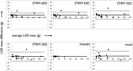
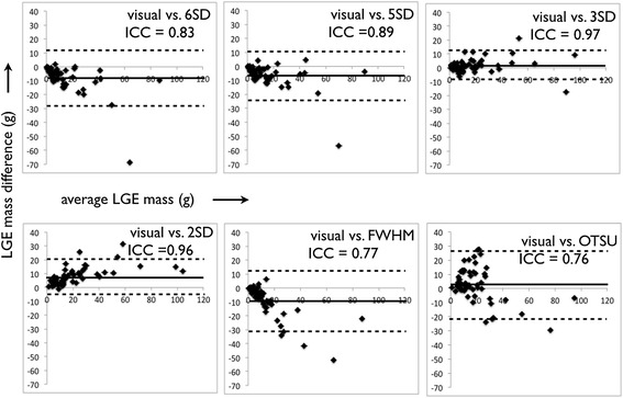
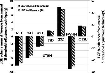
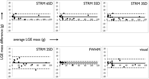
Fifty-two of 76 (68%) patients showed LGE on a total of 202 slices. For accuracy, the STRM >3SD technique showed the greatest agreement with manual segmentation (ICC = 0.97, mean difference and 95% limits of agreement = 1.6 ± 10.7 g) while STRM >6SD, >5SD, 4SD and FWHM techniques systematically underestimated total LGE volume. Slice based analysis of selected SI thresholds similarly showed the STRM >3SD threshold to most closely approximate manually adjusted SI thresholds (ICC = 0.88). For reproducibility, the intra- and inter-observer reproducibility of the >3SD threshold demonstrated an acceptable mean difference and 95% limits of agreement of -0.5 ± 6.8 g and -0.9 ± 5.6 g, respectively.

CONCLUSIONS

FWHM segmentation provides superior reproducibility, however systematically underestimates total LGE volume compared to manual segmentation in patients with HCM. The STRM >3SD technique provides the greatest accuracy while retaining acceptable reproducibility and may therefore be a preferred approach for LGE quantification in this population.

摘要

背景

晚期钆增强(LGE)的存在及范围与肥厚型心肌病(HCM)患者的不良事件相关。信号强度(SI)阈值技术通常用于定量分析;建议半高宽(FWHM)技术比信号阈值与参考均值(STRM)技术具有更高的可重复性,然而,与手动设定最佳SI阈值相比,这些方法的准确性尚未得到研究。在本研究中,我们比较了所有已知的半自动LGE定量技术在HCM患者中的准确性和可重复性。

方法

研究了76例HCM患者(51例男性,年龄54±13岁)。使用7种半自动技术对总LGE体积进行定量,并与专家手动调整SI阈值以实现最佳分割的结果进行比较。测试的技术包括基于STRM的高于参考心肌平均SI的2、3、4、5和6个标准差的阈值、FWHM技术和大津自动阈值(OAT)技术。记录所有切片上每种技术选择的SI阈值。报告每种半自动技术与专家手动调整的LGE分割之间的Bland-Altman分析和组内相关系数(ICC)。还进行了观察者内和观察者间的可重复性评估。

结果

76例患者中有52例(68%)在总共202个切片上显示LGE。在准确性方面,STRM>3SD技术与手动分割的一致性最高(ICC = 0.97,平均差异和95%一致性界限 = 1.6±10.7 g),而STRM>6SD、>5SD、4SD和FWHM技术系统性地低估了总LGE体积。对选定SI阈值的切片分析同样显示,STRM>3SD阈值最接近手动调整的SI阈值(ICC = 0.88)。在可重复性方面,>3SD阈值的观察者内和观察者间可重复性分别显示出可接受的平均差异和95%一致性界限,分别为-0.5±6.8 g和-0.9±5.6 g。

结论

FWHM分割提供了更高的可重复性,然而,与HCM患者的手动分割相比,系统性地低估了总LGE体积。STRM>3SD技术提供了最高的准确性,同时保持了可接受的可重复性,因此可能是该人群LGE定量的首选方法。

https://cdn.ncbi.nlm.nih.gov/pmc/blobs/ed2f/4189726/313cf1b6601c/12968_2014_85_Fig7_HTML.jpg
https://cdn.ncbi.nlm.nih.gov/pmc/blobs/ed2f/4189726/b543f55fb547/12968_2014_85_Fig1_HTML.jpg
https://cdn.ncbi.nlm.nih.gov/pmc/blobs/ed2f/4189726/cdce5bab742f/12968_2014_85_Fig2_HTML.jpg
https://cdn.ncbi.nlm.nih.gov/pmc/blobs/ed2f/4189726/cbdd7e1411af/12968_2014_85_Fig3_HTML.jpg
https://cdn.ncbi.nlm.nih.gov/pmc/blobs/ed2f/4189726/74051d372943/12968_2014_85_Fig4_HTML.jpg
https://cdn.ncbi.nlm.nih.gov/pmc/blobs/ed2f/4189726/16a271d099d1/12968_2014_85_Fig5_HTML.jpg
https://cdn.ncbi.nlm.nih.gov/pmc/blobs/ed2f/4189726/d45b62c0e931/12968_2014_85_Fig6_HTML.jpg
https://cdn.ncbi.nlm.nih.gov/pmc/blobs/ed2f/4189726/313cf1b6601c/12968_2014_85_Fig7_HTML.jpg

相似文献

1
Accuracy and reproducibility of semi-automated late gadolinium enhancement quantification techniques in patients with hypertrophic cardiomyopathy.肥厚型心肌病患者半自动延迟钆增强定量技术的准确性和可重复性
J Cardiovasc Magn Reson. 2014 Oct 7;16(1):85. doi: 10.1186/s12968-014-0085-x.
2
Quantitative analysis of late gadolinium enhancement in hypertrophic cardiomyopathy: comparison of diagnostic performance in myocardial fibrosis between gadobutrol and gadopentetate dimeglumine.肥厚型心肌病延迟钆增强的定量分析:钆布醇与钆喷酸葡胺在心肌纤维化诊断效能上的比较
Int J Cardiovasc Imaging. 2017 Aug;33(8):1191-1200. doi: 10.1007/s10554-017-1101-7. Epub 2017 Mar 13.
3
Comparison of semi-automated scar quantification techniques using high-resolution, 3-dimensional late-gadolinium-enhancement magnetic resonance imaging.使用高分辨率三维延迟钆增强磁共振成像的半自动瘢痕定量技术比较
Int J Cardiovasc Imaging. 2015 Feb;31(2):349-57. doi: 10.1007/s10554-014-0553-2. Epub 2014 Oct 12.
4
Influence of phase correction of late gadolinium enhancement images on scar signal quantification in patients with ischemic and non-ischemic cardiomyopathy.延迟钆增强图像的相位校正对缺血性和非缺血性心肌病患者瘢痕信号定量的影响
J Cardiovasc Magn Reson. 2015 Aug 7;17(1):66. doi: 10.1186/s12968-015-0163-8.
5
Evaluation of techniques for the quantification of myocardial scar of differing etiology using cardiac magnetic resonance.应用心脏磁共振评估不同病因所致心肌瘢痕的量化技术。
JACC Cardiovasc Imaging. 2011 Feb;4(2):150-6. doi: 10.1016/j.jcmg.2010.11.015.
6
Comparison of LGE quantitation methods in cardiac sarcoidosis to predict clinical outcomes.心脏结节病中LGE定量方法对临床结局预测的比较
Int J Cardiovasc Imaging. 2023 Mar;39(3):641-650. doi: 10.1007/s10554-022-02751-4. Epub 2022 Nov 23.
7
Comparison of fast multi-slice and standard segmented techniques for detection of late gadolinium enhancement in ischemic and non-ischemic cardiomyopathy - a prospective clinical cardiovascular magnetic resonance trial.比较快速多层面和标准分段技术在缺血性和非缺血性心肌病中的延迟钆增强检测 - 一项前瞻性临床心血管磁共振试验。
J Cardiovasc Magn Reson. 2018 Feb 19;20(1):13. doi: 10.1186/s12968-018-0434-2.
8
Comparison of semi-automated methods to quantify infarct size and area at risk by cardiovascular magnetic resonance imaging at 1.5T and 3.0T field strengths.在1.5T和3.0T场强下,通过心血管磁共振成像对半自动方法定量梗死面积和危险区域的比较。
BMC Res Notes. 2015 Feb 25;8:52. doi: 10.1186/s13104-015-1007-1.
9
High-resolution 3-dimensional late gadolinium enhancement scar imaging in surgically corrected Tetralogy of Fallot: clinical feasibility of volumetric quantification and visualization.法洛四联症手术矫正后的高分辨率三维延迟钆增强瘢痕成像:容积定量和可视化的临床可行性
J Cardiovasc Magn Reson. 2014 Oct 1;16(1):76. doi: 10.1186/s12968-014-0076-y.
10
Comparison of myocardial fibrosis quantification methods by cardiovascular magnetic resonance imaging for risk stratification of patients with suspected myocarditis.比较心血管磁共振成像心肌纤维化定量方法在疑似心肌炎患者危险分层中的作用。
J Cardiovasc Magn Reson. 2019 Feb 28;21(1):14. doi: 10.1186/s12968-019-0520-0.

引用本文的文献

1
The Role of Cardiac Magnetic Resonance Imaging in the Management of Hypertrophic Cardiomyopathy.心脏磁共振成像在肥厚型心肌病管理中的作用
J Cardiovasc Dev Dis. 2025 May 15;12(5):189. doi: 10.3390/jcdd12050189.
2
Prevalence of Persistent Cardiovascular and Pulmonary Abnormalities on PET/MRI and DECT Imaging in Long COVID Patients.新冠后综合征患者PET/MRI和DECT成像上持续性心血管和肺部异常的患病率
J Nucl Med. 2025 Jul 1;66(7):1126-1134. doi: 10.2967/jnumed.124.268980.
3
Myocardial Fibrosis Quantification Methods by Cardiovascular Magnetic Resonance Imaging in Patients with Fabry Disease.

本文引用的文献

1
Prognostic value of quantitative contrast-enhanced cardiovascular magnetic resonance for the evaluation of sudden death risk in patients with hypertrophic cardiomyopathy.定量对比增强心血管磁共振对肥厚型心肌病患者猝死风险评估的预后价值。
Circulation. 2014 Aug 5;130(6):484-95. doi: 10.1161/CIRCULATIONAHA.113.007094.
2
Role of late gadolinium enhancement cardiovascular magnetic resonance in the risk stratification of hypertrophic cardiomyopathy.延迟钆增强心血管磁共振成像在肥厚型心肌病危险分层中的作用
Heart. 2014 Dec;100(23):1851-8. doi: 10.1136/heartjnl-2013-305471. Epub 2014 Jun 24.
3
Myocardial fibrosis in hypertrophic cardiomyopathy: accurate reflection of histopathological findings by CMR.
法布里病患者心血管磁共振成像心肌纤维化定量方法
J Clin Med. 2024 Aug 26;13(17):5047. doi: 10.3390/jcm13175047.
4
Novel Multiplexed Plasma Biomarker Panel Has Diagnostic and Prognostic Potential in Children With Hypertrophic Cardiomyopathy.新型多重血浆生物标志物panel 在儿童肥厚型心肌病中有诊断和预后潜力。
Circ Genom Precis Med. 2024 Jun;17(3):e004448. doi: 10.1161/CIRCGEN.123.004448. Epub 2024 Jun 7.
5
Evaluation of deep learning-based reconstruction late gadolinium enhancement images for identifying patients with clinically unrecognized myocardial infarction.基于深度学习的重建晚期钆增强图像用于识别临床无症状性心肌梗死患者的评估。
BMC Med Imaging. 2024 May 31;24(1):127. doi: 10.1186/s12880-024-01308-2.
6
Computational modeling of cardiac electrophysiology and arrhythmogenesis: toward clinical translation.心脏电生理学和心律失常发生的计算建模:迈向临床转化。
Physiol Rev. 2024 Jul 1;104(3):1265-1333. doi: 10.1152/physrev.00017.2023. Epub 2023 Dec 28.
7
Quality control-driven deep ensemble for accountable automated segmentation of cardiac magnetic resonance LGE and VNE images.用于心脏磁共振延迟强化(LGE)和心室非增强(VNE)图像可问责自动分割的质量控制驱动深度集成方法。
Front Cardiovasc Med. 2023 Sep 11;10:1213290. doi: 10.3389/fcvm.2023.1213290. eCollection 2023.
8
Systolic anterior motion of the anterior mitral valve leaflet begins in subclinical hypertrophic cardiomyopathy.前二尖瓣瓣叶收缩期前向运动始于亚临床肥厚型心肌病。
Eur Heart J Cardiovasc Imaging. 2023 Dec 21;25(1):86-94. doi: 10.1093/ehjci/jead186.
9
Imaging Predictors of Left Ventricular Functional Recovery after Reperfusion Therapy of ST-Elevation Myocardial Infarction Assessed by Cardiac Magnetic Resonance.通过心脏磁共振评估ST段抬高型心肌梗死再灌注治疗后左心室功能恢复的影像学预测指标
J Cardiovasc Dev Dis. 2023 Jul 11;10(7):294. doi: 10.3390/jcdd10070294.
10
Prognostic Value of Different Thresholds for Myocardial Scar Quantification on Cardiac MRI Late Gadolinium Enhancement Images in Patients Receiving Implantable Cardioverter Defibrillators.植入式心律转复除颤器患者心脏磁共振延迟钆增强图像上不同心肌瘢痕量化阈值的预后价值
Radiol Cardiothorac Imaging. 2023 May 25;5(3):e210247. doi: 10.1148/ryct.210247. eCollection 2023 Jun.
肥厚型心肌病中的心肌纤维化:CMR 对组织病理学发现的准确反映。
JACC Cardiovasc Imaging. 2013 May;6(5):587-96. doi: 10.1016/j.jcmg.2012.09.018. Epub 2013 Apr 10.
4
T1 measurements identify extracellular volume expansion in hypertrophic cardiomyopathy sarcomere mutation carriers with and without left ventricular hypertrophy.T1 测量可识别肥厚型心肌病肌节突变携带者有无左心室肥厚的细胞外容积扩张。
Circ Cardiovasc Imaging. 2013 May 1;6(3):415-22. doi: 10.1161/CIRCIMAGING.112.000333. Epub 2013 Apr 2.
5
Human non-contrast T1 values and correlation with histology in diffuse fibrosis.弥散性纤维化中人类非对比 T1 值与组织学的相关性。
Heart. 2013 Jul;99(13):932-7. doi: 10.1136/heartjnl-2012-303052. Epub 2013 Jan 24.
6
Myocardial tissue characterization using magnetic resonance noncontrast t1 mapping in hypertrophic and dilated cardiomyopathy.应用心脏磁共振钆对比增强 T1 mapping 技术对肥厚型和扩张型心肌病进行心肌组织特征分析。
Circ Cardiovasc Imaging. 2012 Nov;5(6):726-33. doi: 10.1161/CIRCIMAGING.112.976738. Epub 2012 Oct 15.
7
Auto-threshold quantification of late gadolinium enhancement in patients with acute heart disease.自动门控钆延迟增强在急性心脏病患者中的定量分析。
J Magn Reson Imaging. 2013 Feb;37(2):382-90. doi: 10.1002/jmri.23814. Epub 2012 Sep 25.
8
Extracellular volume fraction mapping in the myocardium, part 2: initial clinical experience.心肌细胞外容积分数测绘 2 部分:初步临床经验。
J Cardiovasc Magn Reson. 2012 Sep 11;14(1):64. doi: 10.1186/1532-429X-14-64.
9
Progression of myocardial fibrosis assessed with cardiac magnetic resonance in hypertrophic cardiomyopathy.肥厚型心肌病中心肌纤维化的心脏磁共振评估。
J Am Coll Cardiol. 2012 Sep 4;60(10):922-9. doi: 10.1016/j.jacc.2012.03.076.
10
Intermediate-signal-intensity late gadolinium enhancement predicts ventricular tachyarrhythmias in patients with hypertrophic cardiomyopathy.延迟钆增强中期信号强度可预测肥厚型心肌病患者的室性心动过速/心搏过速。
Circ Cardiovasc Imaging. 2012 Jan;5(1):78-85. doi: 10.1161/CIRCIMAGING.111.963819. Epub 2011 Dec 1.