Mo Juan, Zhang Ming, Marshall Brendan, Smith Sylvia, Covar Jason, Atherton Sally
Georgia Regents University, Medical College of Georgia, Department of Cellular Biology and Anatomy, Augusta, GA.
Georgia Regents University, Medical College of Georgia, Department of Cellular Biology and Anatomy, Augusta, GA ; Georgia Regents University, Medical College of Georgia, Department of Ophthalmology, Augusta, GA.
Mol Vis. 2014 Aug 14;20:1161-73. eCollection 2014.
Previous studies have demonstrated that autophagy is involved in the pathogenesis of human cytomegalovirus (HCMV) infection. However, whether autophagy is regulated by murine cytomegalovirus (MCMV) infection has not yet been investigated. The purpose of these studies was to determine how autophagy is affected by MCMV infection of the retinal pigment epithelial (RPE) cells and whether there is a functional relationship between autophagy and apoptosis; and if so, how regulation of autophagy impacts apoptosis.
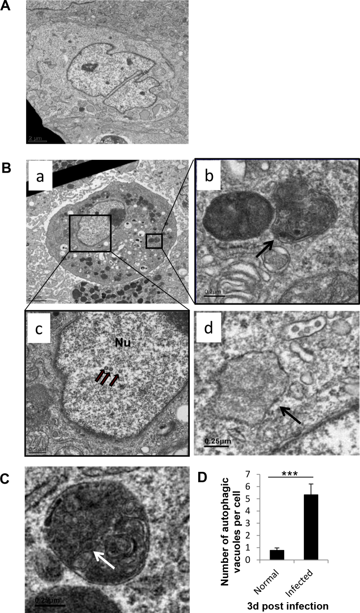
RPE cells were isolated from C57BL/6 mice and infected with MCMV K181. The cells were cultured in medium containing rapamycin, chloroquine, or ammonium chloride. Green fluorescent protein-light chain 3 (GFP-LC3) plasmid was transfected to RPE cells, and the GFP-LC3 positive puncta were counted. Electron microscopic (EM) images were taken to visualize the structure of the autophagic vacuoles. Western blot was performed to detect the expression of related proteins. Trypan blue exclusion assay was used to measure the percentage of viable cells.
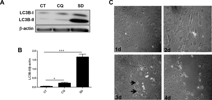
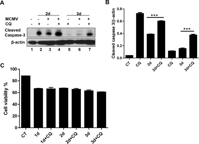
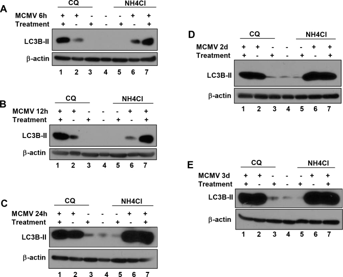
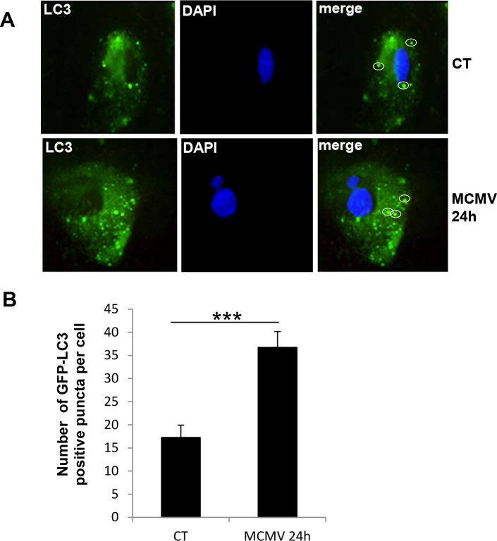
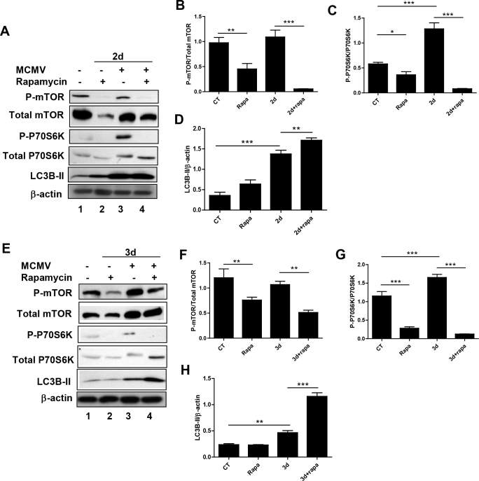
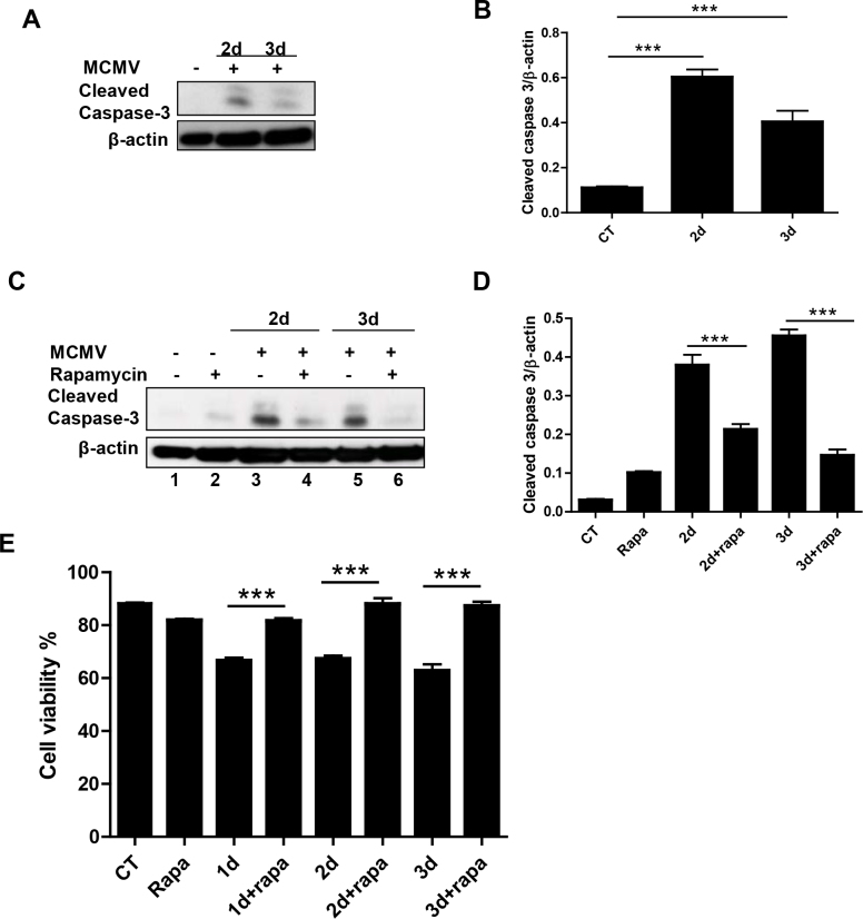
Although the LC3B-II levels consistently increased during MCMV infection of RPE cells, administration of chloroquine or ammonium chloride increased LC3B-II expression only at the early stage of infection (6 h post-inoculation [p.i.] and 12 h p.i.), not at or after 24 h p.i. The punctate autophagic vacuoles in the GFP-LC3 transfected RPE cells were counted using light microscopy or by EM examination, the number of autophagic vacuoles was significantly increased in the MCMV-infected RPE cells compared to the uninfected controls. Compared to untreated MCMV-infected control cells, rapamycin treatment resulted in a significant decrease in the cleaved caspase 3 levels as well as a significant decrease in the ratio of phosphorylated mammalian target of rapamycin (mTOR) to total mTOR and in the ratio of phosphorylated P70S6K to total P70S6K. In contrast, chloroquine treatment resulted in a significant increase in the cleaved caspase 3 levels in the MCMV-infected RPE cells.
Autophagic vacuole accumulation was detected during MCMV infection of RPE cells. In contrast, autophagic flux was greatly decreased at or after 24 h p.i. The results suggest that MCMV might have a strategy for inhibiting or blocking autophagy activity by targeting a later autophagy process, such as the formation of autolysosomes or degradation of their content. Our data also suggest that there is a functional relationship between autophagy and apoptosis, which plays an important role during MCMV infection of the RPE.
先前的研究表明自噬参与人巨细胞病毒(HCMV)感染的发病机制。然而,小鼠巨细胞病毒(MCMV)感染是否调节自噬尚未得到研究。这些研究的目的是确定视网膜色素上皮(RPE)细胞的MCMV感染如何影响自噬,以及自噬与凋亡之间是否存在功能关系;如果存在,自噬的调节如何影响凋亡。
从C57BL/6小鼠分离RPE细胞,并感染MCMV K181。将细胞培养在含有雷帕霉素、氯喹或氯化铵的培养基中。将绿色荧光蛋白-轻链3(GFP-LC3)质粒转染到RPE细胞中,并对GFP-LC3阳性斑点进行计数。拍摄电子显微镜(EM)图像以观察自噬泡的结构。进行蛋白质印迹法检测相关蛋白的表达。使用台盼蓝排斥试验测量活细胞百分比。
尽管在RPE细胞的MCMV感染期间LC3B-II水平持续升高,但给予氯喹或氯化铵仅在感染早期(接种后6小时[p.i.]和12小时p.i.)增加LC3B-II表达,在24小时p.i.时或之后则没有增加。使用光学显微镜或通过EM检查对GFP-LC3转染的RPE细胞中的点状自噬泡进行计数,与未感染的对照相比,MCMV感染的RPE细胞中自噬泡的数量显著增加。与未处理的MCMV感染对照细胞相比,雷帕霉素处理导致裂解的半胱天冬酶3水平显著降低,以及磷酸化的雷帕霉素哺乳动物靶标(mTOR)与总mTOR的比率以及磷酸化的P70S6K与总P70S6K的比率显著降低。相反,氯喹处理导致MCMV感染的RPE细胞中裂解的半胱天冬酶3水平显著增加。
在RPE细胞的MCMV感染期间检测到自噬泡积累。相反,在24小时p.i.时或之后自噬通量大大降低。结果表明,MCMV可能有一种通过靶向后期自噬过程(如自溶酶体的形成或其内容物的降解)来抑制或阻断自噬活性的策略。我们的数据还表明自噬与凋亡之间存在功能关系,这在RPE细胞的MCMV感染期间起重要作用。