Agladioglu Kadir, Ardic Fazıl Necdet, Tumkaya Funda, Bir Ferda
Department of Radiology, Pamukkale University, Denizli, Turkey.
Department of Otolaryngology Head and Neck Surgery, Pamukkale University, Denizli, Turkey.
Pol J Radiol. 2014 Oct 13;79:360-2. doi: 10.12659/PJR.890795. eCollection 2014.
Intrasphenoidal encephalocele (ISE) is a rare clinical entity. The incidence of congenital encephalocele is very low. Accurate diagnosis and surgical approach is of critical value.
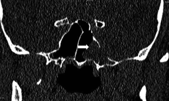
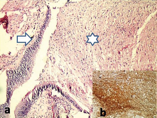
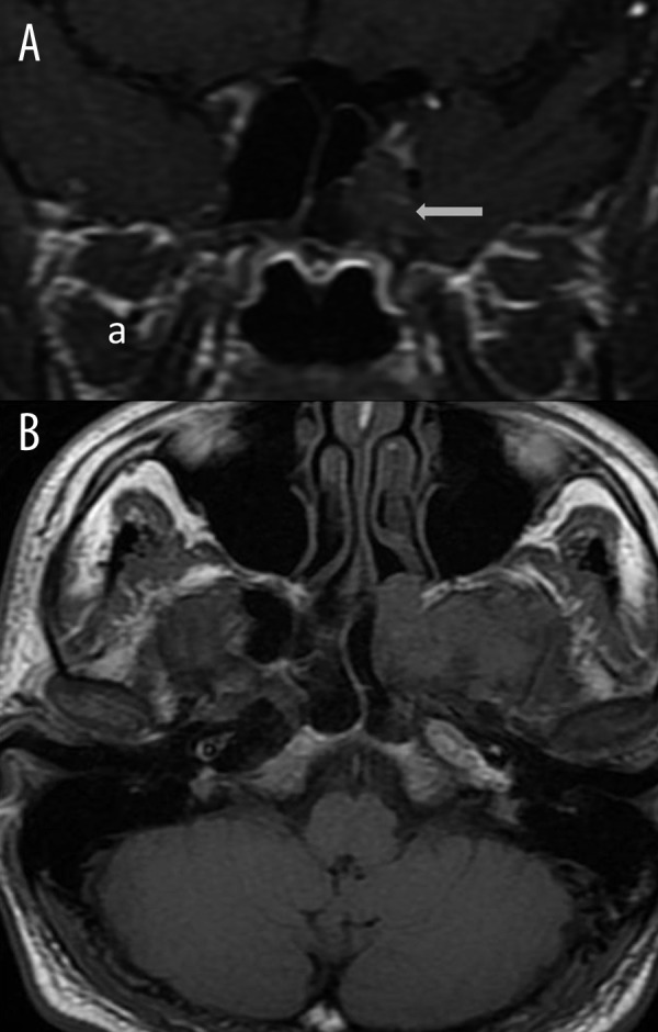
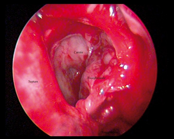
We present a case of intrasphenoidal encephalocele in a 40-year-old man. He complained of cerebrospinal fluid (CSF) rhinorrhea and recurrent meningitis. In images of computed tomography (CT) and magnetic resonance imaging (MRI), intrasphenoidal encephalocele herniating through a defect of the left lateral sphenoid sinus wall was determined. Incisional biopsies were taken by endoscopic transnasal approach and histopathological examination revealed an encephalocele. In the differential diagnosis, ISE can be taken for inflammatory or malignant sinusoidal soft tissue masses. ISE is differentiated from other entities by demonstrating continuity with normal brain tissue.
MRI clearly demonstrates that the herniating soft tissue is isointense with brain and continuous with brain tissue via the sphenoid sinus, thereby the treatment decision-making process is very important.
蝶窦内脑膨出(ISE)是一种罕见的临床病症。先天性脑膨出的发病率很低。准确的诊断和手术方法具有关键价值。
我们报告一例40岁男性的蝶窦内脑膨出病例。他主诉脑脊液鼻漏和复发性脑膜炎。在计算机断层扫描(CT)和磁共振成像(MRI)图像中,确定蝶窦内脑膨出通过左侧蝶窦壁的缺损疝出。通过鼻内镜经鼻途径进行切开活检,组织病理学检查显示为脑膨出。在鉴别诊断中,ISE可能被误诊为炎性或恶性鼻窦软组织肿块。通过证明与正常脑组织的连续性,ISE可与其他病症相鉴别。
MRI清楚地显示疝出的软组织与脑等信号,并通过蝶窦与脑组织连续,因此治疗决策过程非常重要。