Ngoc Doan-Van, Trung Nguyen Ngoc, Duc Le Anh, Sang Nguyen-Van, Ninh Tran Phan, My Thieu-Thi Tra, Duc Nguyen Minh
Department of Medical Imaging Technology, VNU University of Medicine and Pharmacy, Hanoi, Vietnam.
Department of Radiology, E hospital, Ha Noi, Vietnam.
Radiol Case Rep. 2021 Aug 3;16(10):2945-2948. doi: 10.1016/j.radcr.2021.06.064. eCollection 2021 Oct.
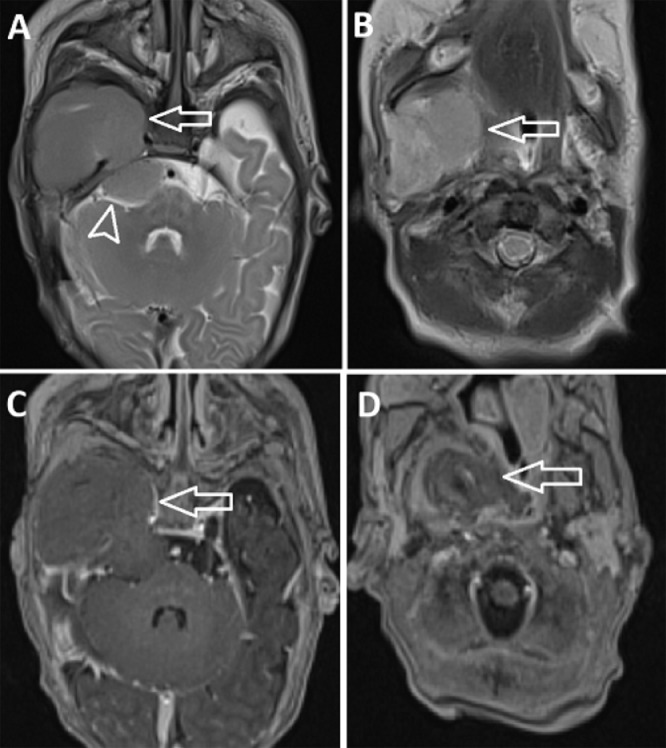
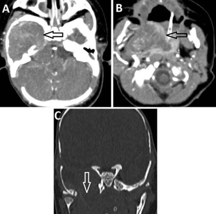
An encephalocele may be congenital or acquired and is characterized by the herniation of cranial contents through a skull bone defect. Most congenital encephaloceles occur in the occipital area, and temporal bone encephaloceles in children are rare. Congenital encephaloceles can be diagnosed either prenatally or after birth. We describe the case of a congenital temporal bone encephalocele in a 2-month-old boy that was diagnosed after birth. The patient presented with seizures and a bulging mass in the right neck that was detected by his mother during the second month after birth. The combined results from brain magnetic resonance imaging, computed tomography, and histological analysis confirmed the diagnosis of encephalocele. Although the surgical repair was offered, the family declined.
脑膨出可分为先天性或后天性,其特征是颅骨内容物通过颅骨骨缺损处疝出。大多数先天性脑膨出发生在枕部区域,儿童颞骨脑膨出较为罕见。先天性脑膨出可在产前或出生后诊断。我们描述了一名2个月大男婴先天性颞骨脑膨出的病例,该病例在出生后被诊断出来。患儿出生后第二个月,其母亲发现患儿出现癫痫发作,右颈部有一肿物隆起。脑磁共振成像、计算机断层扫描和组织学分析的综合结果证实了脑膨出的诊断。尽管提供了手术修复方案,但家属拒绝了。