Ibrahim Mohamed Magdy, Bond Jennifer, Bergeron Andrew, Miller Kyle J, Ehanire Tosan, Quiles Carlos, Lorden Elizabeth R, Medina Manuel A, Fisher Mark, Klitzman Bruce, Selim M Angelica, Leong Kam W, Levinson Howard
Division of Plastic and Reconstructive Surgery, Department of Surgery, Duke University School of Medicine, Durham, North Carolina.
Wound Repair Regen. 2014 Nov-Dec;22(6):755-64. doi: 10.1111/wrr.12238. Epub 2015 Jan 8.
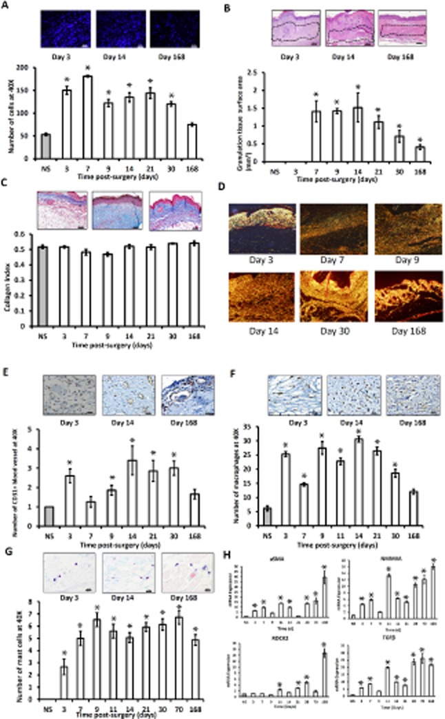
Hypertrophic scar (HSc) contraction following burn injury causes contractures. Contractures are painful and disfiguring. Current therapies are marginally effective. To study pathogenesis and develop new therapies, a murine model is needed. We have created a validated immune-competent murine HSc model. A third-degree burn was created on dorsum of C57BL/6 mice. Three days postburn, tissue was excised and grafted with ear skin. Graft contraction was analyzed and tissue harvested on different time points. Outcomes were compared with human condition to validate the model. To confirm graft survival, green fluorescent protein (GFP) mice were used, and histologic analysis was performed to differentiate between ear and back skin. Role of panniculus carnosus in contraction was analyzed. Cellularity was assessed with 4',6-diamidino-2-phenylindole. Collagen maturation was assessed with Picro-sirius red. Mast cells were stained with Toluidine blue. Macrophages were detected with F4/80 immune. Vascularity was assessed with CD31 immune. RNA for contractile proteins was detected by quantitative real-time polymerase chain reaction (qRT-PCR). Elastic moduli of skin and scar tissue were analyzed using a microstrain analyzer. Grafts contracted to ∼45% of their original size by day 14 and maintained their size. Grafting of GFP mouse skin onto wild-type mice, and analysis of dermal thickness and hair follicle density, confirmed graft survival. Interestingly, hair follicles disappeared after grafting and regenerated in ear skin configuration by day 30. Radiological analysis revealed that panniculus carnosus doesn't contribute to contraction. Microscopic analyses showed that grafts show increase in cellularity. Granulation tissue formed after day 3. Collagen analysis revealed increases in collagen maturation over time. CD31 stain revealed increased vascularity. Macrophages and mast cells were increased. qRT-PCR showed up-regulation of transforming growth factor beta, alpha smooth muscle actin, and rho-associated protein kinase 2 in HSc. Tensile testing revealed that human skin and scar tissues are tougher than mouse skin and scar tissues.
烧伤后肥厚性瘢痕(HSc)收缩会导致挛缩。挛缩会引起疼痛且影响外观。目前的治疗方法效果甚微。为了研究发病机制并开发新的治疗方法,需要一个小鼠模型。我们创建了一个经过验证的具有免疫活性的小鼠HSc模型。在C57BL/6小鼠的背部制造三度烧伤。烧伤后三天,切除组织并用耳部皮肤进行移植。分析移植组织的收缩情况,并在不同时间点采集组织。将结果与人类情况进行比较以验证该模型。为了确认移植组织的存活情况,使用了绿色荧光蛋白(GFP)小鼠,并进行组织学分析以区分耳部皮肤和背部皮肤。分析了肉膜在收缩中的作用。用4',6-二脒基-2-苯基吲哚评估细胞数量。用苦味酸天狼星红评估胶原蛋白成熟度。用甲苯胺蓝对肥大细胞进行染色。用F4/80免疫法检测巨噬细胞。用CD31免疫法评估血管生成。通过定量实时聚合酶链反应(qRT-PCR)检测收缩蛋白的RNA。使用微应变分析仪分析皮肤和瘢痕组织的弹性模量。移植组织在第14天时收缩至其原始大小的约45%,并保持其大小。将GFP小鼠皮肤移植到野生型小鼠上,并分析真皮厚度和毛囊密度,证实了移植组织的存活。有趣的是,移植后毛囊消失,并在第30天时以耳部皮肤的形态再生。放射学分析表明肉膜对收缩没有作用。显微镜分析显示移植组织的细胞数量增加。第3天后形成肉芽组织。胶原蛋白分析显示随着时间的推移胶原蛋白成熟度增加。CD31染色显示血管生成增加。巨噬细胞和肥大细胞数量增加。qRT-PCR显示HSc中转化生长因子β、α平滑肌肌动蛋白和Rho相关蛋白激酶2上调。拉伸试验表明人类皮肤和瘢痕组织比小鼠皮肤和瘢痕组织更坚韧。