Institute for Biological and Medical Imaging, Helmholtz Zentrum München, Ingolstädter Landstr. 1, 85764 Neuherberg, Germany ; Chair for Biological Imaging, Technische Universität München, Ismaninger Str. 22, 81675 München, Germany.
Department of Radiology, Klinikum Rechts der Isar, Technische Universität München, Ismaninger Str. 22, 81675 München, Germany.
Photoacoustics. 2012 Dec 10;1(1):3-8. doi: 10.1016/j.pacs.2012.11.001. eCollection 2013 Mar.
To investigate the feasibility of a high resolution optical imaging strategy for myocardial infarction.
Near-infrared approaches to imaging cardiovascular disease enable visualization of disease-associated biological processes in vivo. However, even at the scale of small animals, the strong scattering of light prevents high resolution imaging after the first 1-2 mm of tissue, leading to degraded signal localization.
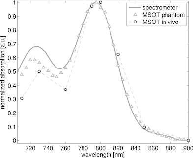
Multispectral optoacoustic tomography (MSOT) was used to non-invasively image myocardial infarction (MI) in a murine model of coronary artery ligation at resolutions not possible with current deep-tissue optical imaging methods. Post-MI imaging was based on resolving the spectral absorption signature of a dendritic polyglycerol sulfate-based (dPGS) near-infrared imaging agent targeted to P- and L-selectin.
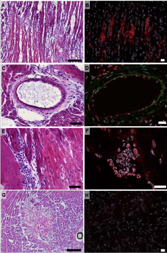
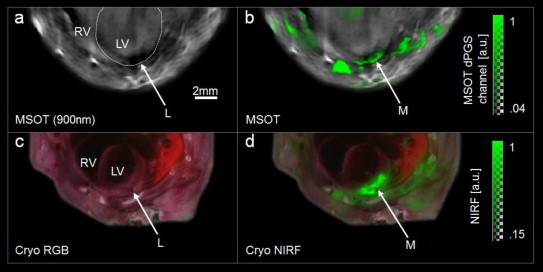
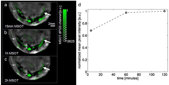
In vivo imaging succeeded in detection of the agent in the injured myocardium after intravenous injection. The high anatomic resolution (<200 μm) achieved by the described method allowed signals originating in the infarcted heart to be distinguished from uptake in adjacent regions. Histological analysis found dPGS signal in infarcted areas, originating from leukocytes and endothelial cells.
MSOT imaging of myocardial infarction provides non-invasive visualization of optical contrast with a high spatial resolution that is not degraded by the scattering of light.
探索一种用于心肌梗死的高分辨率光学成像策略的可行性。
近红外方法可用于心血管疾病成像,使我们能够在体内可视化与疾病相关的生物过程。然而,即使在小动物的范围内,光的强散射也会阻止在组织最初的 1-2 毫米之后进行高分辨率成像,从而导致信号定位恶化。
多光谱光声断层扫描(MSOT)用于在冠状动脉结扎的小鼠模型中对心肌梗死(MI)进行非侵入性成像,其分辨率无法通过当前的深层组织光学成像方法实现。MI 后成像基于解析针对 P 选择素和 L 选择素的树枝状多聚甘油硫酸酯(dPGS)近红外成像剂的光谱吸收特征。
静脉注射后,体内成像成功地在受损心肌中检测到了该试剂。该方法实现的高解剖分辨率(<200 μm)允许区分起源于梗死心脏的信号与相邻区域的摄取。组织学分析发现 dPGS 信号存在于梗死区域,源自白细胞和内皮细胞。
心肌梗死的 MSOT 成像提供了非侵入性可视化,具有不受光散射影响的高空间分辨率的光学对比度。