Zhang Ning, Chu Eagle S H, Zhang Jingwan, Li Xiaoxing, Liang Qiaoyi, Chen Jie, Chen Minhu, Teoh Narci, Farrell Geoffrey, Sung Joseph J Y, Yu Jun
Institute of Digestive Disease and Department of Medicine and Therapeutics, State Key Laboratory of Digestive Disease and Li Ka Shing Institute of Health Sciences, The Chinese University of Hong Kong, Hong Kong SAR, China. Department of Gastroenterology, First Affiliated Hospital, Sun Yat-sen University, Guangzhou, China.
Institute of Digestive Disease and Department of Medicine and Therapeutics, State Key Laboratory of Digestive Disease and Li Ka Shing Institute of Health Sciences, The Chinese University of Hong Kong, Hong Kong SAR, China. Gastrointestinal Cancer Biology & Therapeutics Laboratory, CUHK-Shenzhen Research Institute, Shenzhen, China.
Oncotarget. 2014 Sep 30;5(18):8330-40. doi: 10.18632/oncotarget.2212.
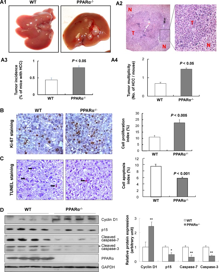
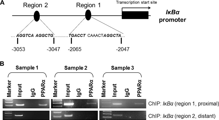
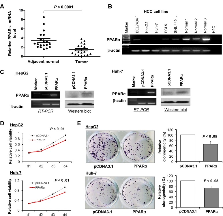
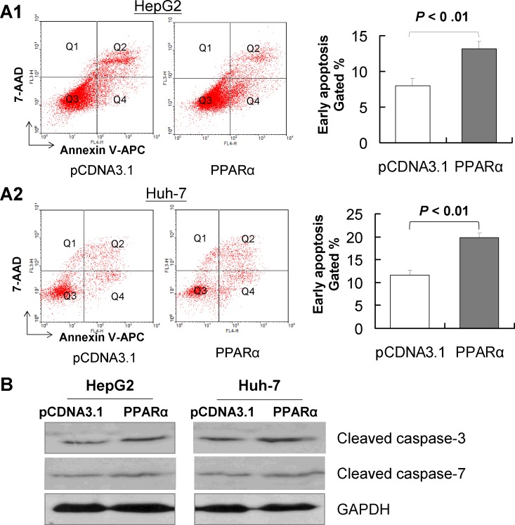
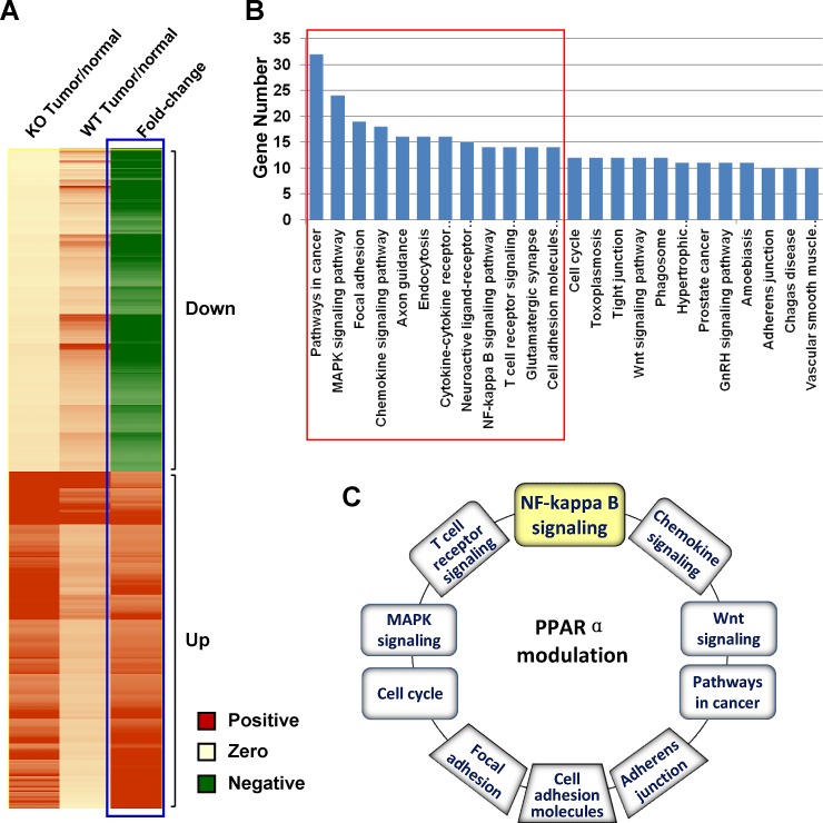
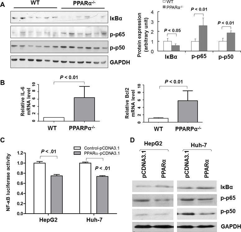
Peroxisome proliferator-activated receptor alpha (PPARα) ligands have been reported to suppress cancer growth. However, the role of PPARα in hepatocarcinogenesis remains unclear. We investigated the functional significance of PPARα in HCC. PPARα-knockout (PPARα-/-) mice were more susceptible to diethylnitrosamine (DEN)-induced HCC at 6 months compared with wild-type (WT) littermates (80% versus 43%, P < 0.05). In resected HCCs, TUNEL-positive apoptotic cells were significantly less in PPARα-/- mice than in WT mice (P < 0.01), commensurate with a reduction in cleaved caspase-3 and caspase-7 protein expression. Ki-67 staining showed increased cell proliferation in PPARα-/- mice (P < 0.01), with concomitant up-regulation of cyclin-D1 and down-regulation of p15. Moreover, ectopic expression of PPARα in HCC cells significantly suppressed cell proliferation and induced apoptosis. The anti-tumorigenic function of PPARα was mediated via NF-κB as evidenced by inhibition of NF-κB promoter activity, diminution of phosphor-p65, phosphor-p50 and BCL2 levels, and enhancing IkBα protein. Chromatin immunoprecipitation analysis confirmed PPARαdirectly binds to the IkBα promoter. In conclusion, PPARα deficiency enhances susceptibility to DEN-initiated HCC. PPARα suppresses tumor cell growth by inhibiting cell proliferation and inducing cell apoptosis via direct targeting IκBα and NF-κB signaling pathway.
据报道,过氧化物酶体增殖物激活受体α(PPARα)配体可抑制癌症生长。然而,PPARα在肝癌发生中的作用仍不清楚。我们研究了PPARα在肝癌中的功能意义。与野生型(WT)同窝小鼠相比,PPARα基因敲除(PPARα-/-)小鼠在6个月时对二乙基亚硝胺(DEN)诱导的肝癌更敏感(80%对43%,P<0.05)。在切除的肝癌组织中,PPARα-/-小鼠的TUNEL阳性凋亡细胞明显少于WT小鼠(P<0.01),这与裂解的半胱天冬酶-3和半胱天冬酶-7蛋白表达的降低相一致。Ki-67染色显示PPARα-/-小鼠的细胞增殖增加(P<0.01),同时细胞周期蛋白D1上调,p15下调。此外,在肝癌细胞中异位表达PPARα可显著抑制细胞增殖并诱导凋亡。PPARα的抗肿瘤功能是通过NF-κB介导的,这表现为NF-κB启动子活性受到抑制、磷酸化p65、磷酸化p50和BCL2水平降低以及IkBα蛋白增加。染色质免疫沉淀分析证实PPARα直接结合到IkBα启动子上。总之,PPARα缺乏会增加对DEN引发的肝癌的易感性。PPARα通过直接靶向IκBα和NF-κB信号通路抑制细胞增殖并诱导细胞凋亡,从而抑制肿瘤细胞生长。