Schlaubitz Silke, Derkaoui Sidi Mohammed, Marosa Lydia, Miraux Sylvain, Renard Martine, Catros Sylvain, Le Visage Catherine, Letourneur Didier, Amédée Joëlle, Fricain Jean-Christophe
CIC 1401, University hospital of Bordeaux/Inserm, Bordeaux, France.
U1148, LVTS/Inserm, Paris, France; Près Sorbonne Paris Cité, University of Paris Nord and University Paris Diderot, Paris, France.
PLoS One. 2014 Oct 20;9(10):e110251. doi: 10.1371/journal.pone.0110251. eCollection 2014.
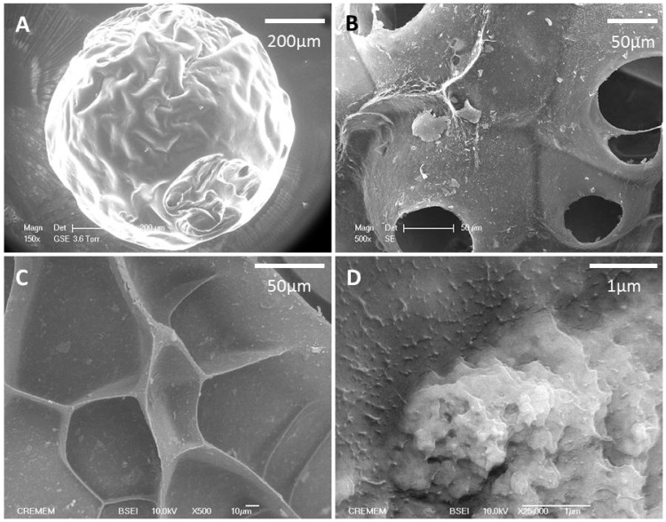
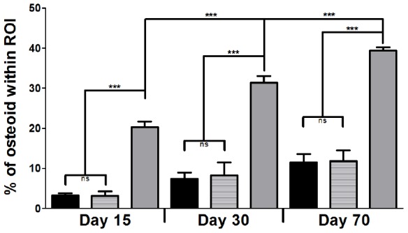
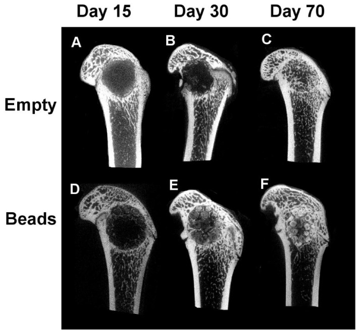
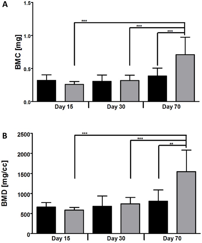
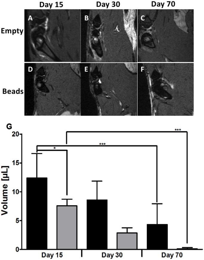
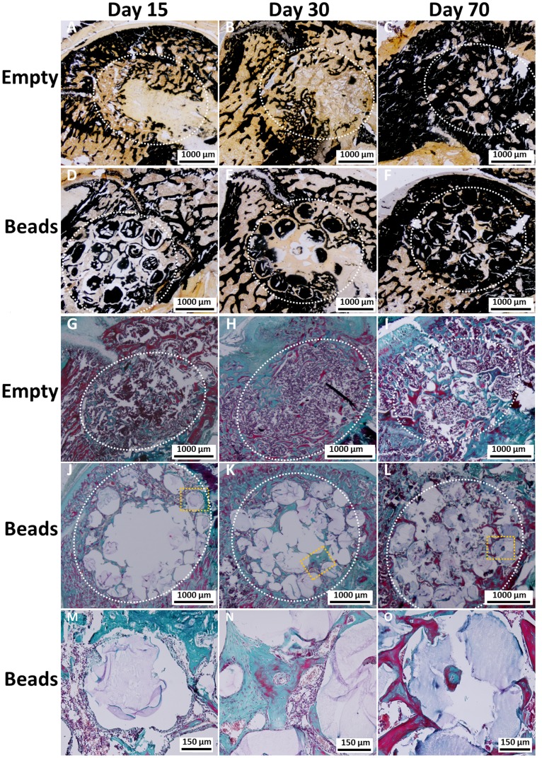
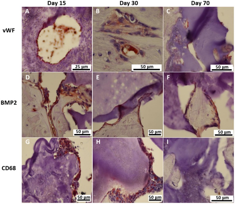
The repair of bone defects is of particular interest for orthopedic, oral, maxillofacial, and dental surgery. Bone loss requiring reconstruction is conventionally addressed through bone grafting. Depending on the size and the location of the defect, this method has limits and risks. Biomaterials can offer an alternative and have features supporting bone repair. Here, we propose to evaluate the cellular penetration and bone formation of new macroporous beads based on pullulan/dextran that has been supplemented with nanocrystalline hydroxyapatite in a rat model. Cross-linked beads of 300-500 µm diameters were used in a lateral femoral condyle defect and analyzed by magnetic resonance imaging, micro-computed tomography, and histology in comparison to the empty defects 15, 30, and 70 days after implantation. Inflammation was absent for both conditions. For empty defects, cellularisation and mineralization started from the periphery of the defect. For the defects containing beads, cellular structures filling out the spaces between the scaffolds with increasing interconnectivity and trabecular-like organization were observed over time. The analysis of calcified sections showed increased mineralization over time for both conditions, but was more pronounced for the samples containing beads. Bone Mineral Density and Bone Mineral Content were both significantly higher at day 70 for the beads in comparison to empty defects as well as compared with earlier time points. Analysis of newly formed tissue around the beads showed an increase of osteoid tissue, measured as percentage of the defect surface. This study suggests that the use of beads for the repair of small size defects in bone may be expanded on to meet the clinical need for a ready-to-use fill-up material that can favor bone formation and mineralization, as well as promote vessel ingrowth into the defect site.
骨缺损的修复在骨科、口腔、颌面和牙科手术中备受关注。传统上,需要重建的骨丢失通过骨移植来解决。根据缺损的大小和位置,这种方法存在局限性和风险。生物材料可提供一种替代方案,并具有支持骨修复的特性。在此,我们提议在大鼠模型中评估基于支链淀粉/葡聚糖并添加了纳米晶羟基磷灰石的新型大孔微珠的细胞穿透和骨形成情况。将直径为300 - 500 µm的交联微珠用于股骨外侧髁缺损,并在植入后15、30和70天通过磁共振成像、微计算机断层扫描和组织学与空白缺损进行比较分析。两种情况均未出现炎症。对于空白缺损,细胞化和矿化从缺损周边开始。对于含有微珠的缺损,随着时间推移,观察到细胞结构填充支架之间的空间,连通性增加且呈小梁样组织。钙化切片分析显示,两种情况的矿化均随时间增加,但含有微珠的样本更为明显。与空白缺损以及早期时间点相比,微珠在第70天时的骨密度和骨矿物质含量均显著更高。对微珠周围新形成组织的分析显示,类骨质组织增加,以缺损表面百分比衡量。本研究表明,用于修复小尺寸骨缺损的微珠应用范围可能会扩大,以满足临床对一种现成的填充材料的需求,该材料可促进骨形成和矿化,并促进血管长入缺损部位。