Department of Neurobiology, Care Sciences and Society, Karolinska Institutet Stockholm, Sweden.
Department of Neuroimaging, Institute of Psychiatry, King's College London London, UK ; NIHR Biomedical Research Centre for Mental Health and NIHR Biomedical Research Unit for Dementia London, UK.
Front Aging Neurosci. 2014 Oct 7;6:264. doi: 10.3389/fnagi.2014.00264. eCollection 2014.
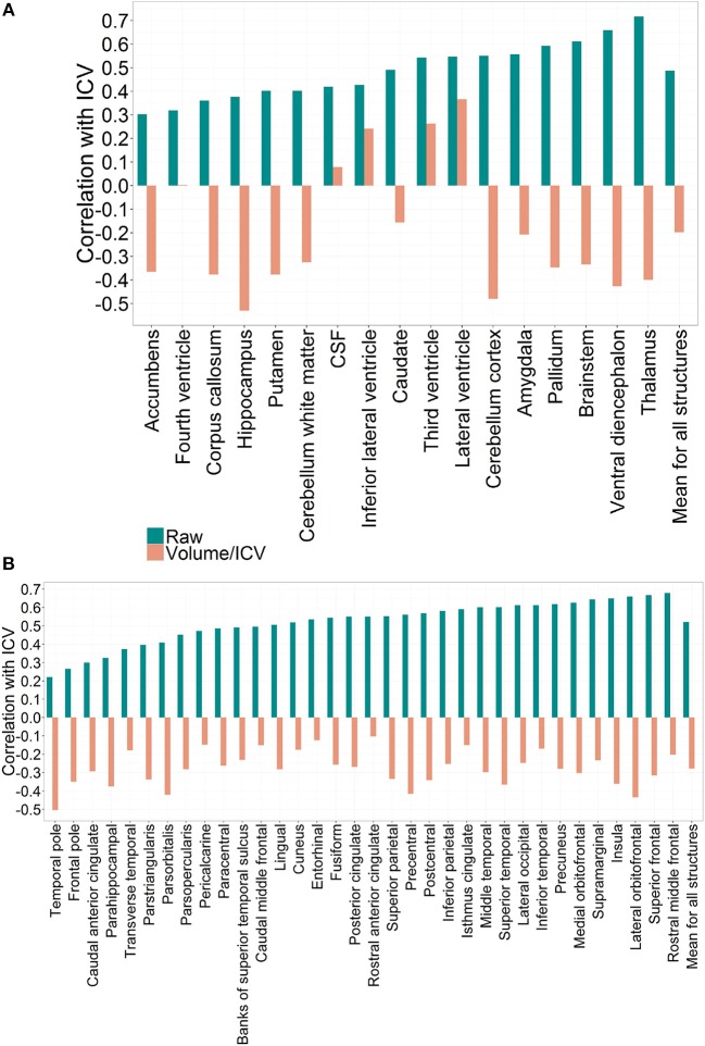
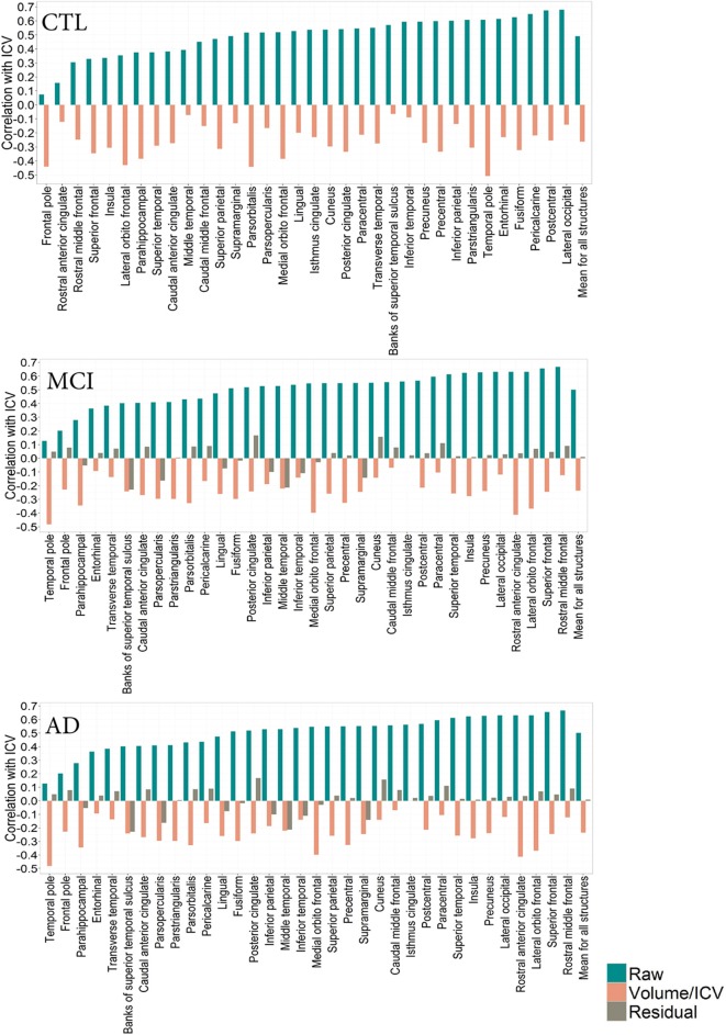
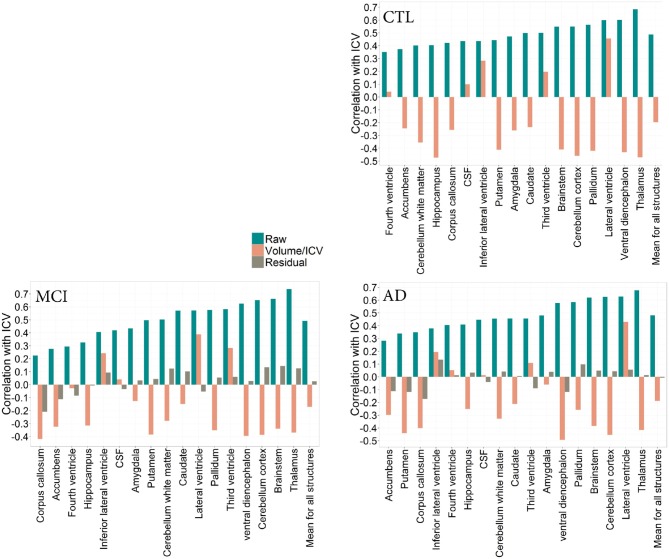
In neurodegeneration research, normalization of regional volumes by intracranial volume (ICV) is important to estimate the extent of disease-driven atrophy. There is little agreement as to whether raw volumes, volume-to-ICV fractions or regional volumes from which the ICV factor has been regressed out should be used for volumetric brain imaging studies. Using multiple regional cortical and subcortical volumetric measures generated by Freesurfer (51 in total), the main aim of this study was to elucidate the implications of these adjustment approaches. Magnetic resonance imaging (MRI) data were analyzed from two large cohorts, the population-based PIVUS cohort (N = 406, all subjects age 75) and the Alzheimer disease Neuroimaging Initiative (ADNI) cohort (N = 724). Further, we studied whether the chosen ICV normalization approach influenced the relationship between hippocampus and cognition in the three diagnostic groups of the ADNI cohort (Alzheimer's disease, mild cognitive impairment, and healthy individuals). The ability of raw vs. adjusted hippocampal volumes to predict diagnostic status was also assessed. In both cohorts raw volumes correlate positively with ICV, but do not scale directly proportionally with it. The correlation direction is reversed for all volume-to-ICV fractions, except the lateral and third ventricles. Most gray matter fractions are larger in females, while lateral ventricle fractions are greater in males. Residual correction effectively eliminated the correlation between the regional volumes and ICV and removed gender differences. The association between hippocampal volumes and cognition was not altered by ICV normalization. Comparing prediction of diagnostic status using the different approaches, small but significant differences were found. The choice of normalization approach should be carefully considered when designing a volumetric brain imaging study.
在神经退行性疾病研究中,通过颅内体积(ICV)对区域体积进行标准化对于估计疾病驱动性萎缩的程度非常重要。对于应该使用原始体积、体积与 ICV 的分数还是从 ICV 因子中回归的区域体积来进行容积脑成像研究,目前尚无共识。本研究使用 Freesurfer 生成的多个皮质和皮质下区域容积测量值(总共 51 个),主要目的是阐明这些调整方法的意义。对来自两个大型队列(基于人群的 PIVUS 队列(N=406,所有受试者年龄均为 75 岁)和阿尔茨海默病神经影像学倡议(ADNI)队列(N=724)的磁共振成像(MRI)数据进行了分析。此外,我们还研究了所选的 ICV 归一化方法是否会影响 ADNI 队列中三个诊断组(阿尔茨海默病、轻度认知障碍和健康个体)中海马体与认知之间的关系。还评估了原始和调整后的海马体体积是否可以预测诊断状态。在两个队列中,原始体积与 ICV 呈正相关,但与 ICV 不成直接比例缩放。除了侧脑室和第三脑室之外,所有体积与 ICV 的分数的相关性方向均相反。大多数灰质分数在女性中更大,而侧脑室分数在男性中更大。残差校正有效地消除了区域体积与 ICV 之间的相关性,并消除了性别差异。海马体体积与认知之间的关联未因 ICV 归一化而改变。使用不同方法比较诊断状态的预测,发现存在微小但显著的差异。在设计容积脑成像研究时,应仔细考虑归一化方法的选择。