• 文献检索
  • 文档翻译
  • 深度研究
  • 学术资讯
  • Suppr Zotero 插件Zotero 插件
  • 邀请有礼
  • 套餐&价格
  • 历史记录
应用&插件
Suppr Zotero 插件Zotero 插件浏览器插件Mac 客户端Windows 客户端微信小程序
定价
高级版会员购买积分包购买API积分包
服务
文献检索文档翻译深度研究API 文档MCP 服务
关于我们
关于 Suppr公司介绍联系我们用户协议隐私条款
关注我们

Suppr 超能文献

核心技术专利:CN118964589B侵权必究
粤ICP备2023148730 号-1Suppr @ 2026

文献检索

告别复杂PubMed语法,用中文像聊天一样搜索,搜遍4000万医学文献。AI智能推荐,让科研检索更轻松。

立即免费搜索

文件翻译

保留排版,准确专业,支持PDF/Word/PPT等文件格式,支持 12+语言互译。

免费翻译文档

深度研究

AI帮你快速写综述,25分钟生成高质量综述,智能提取关键信息,辅助科研写作。

立即免费体验

在7T磁场下测量肾组织弛豫时间。

Measuring renal tissue relaxation times at 7 T.

作者信息

Li Xiufeng, Bolan Patrick J, Ugurbil Kamil, Metzger Gregory J

机构信息

Center for Magnetic Resonance Research, University of Minnesota, Minneapolis, MN, USA.

出版信息

NMR Biomed. 2015 Jan;28(1):63-9. doi: 10.1002/nbm.3195. Epub 2014 Oct 23.

DOI:10.1002/nbm.3195
PMID:25346367
原文链接:https://pmc.ncbi.nlm.nih.gov/articles/PMC4335643/
Abstract

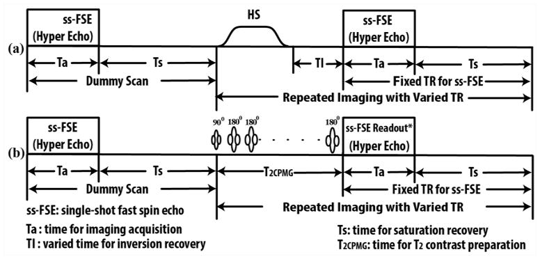
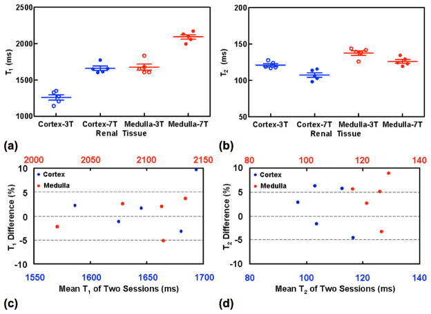
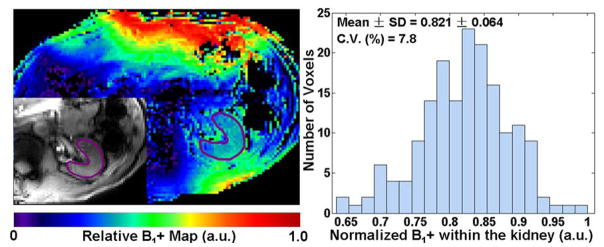
As developments in RF coils and RF management strategies make performing ultra-high-field renal imaging feasible, understanding the relaxation times of the tissue becomes increasingly important for tissue characterization, sequence optimization and quantitative functional renal imaging, such as renal perfusion imaging using arterial spin labeling. By using a magnetization-prepared single-breath-hold fast spin echo imaging method, human renal T1 and T2 imaging studies were successfully performed at 7 T with 11 healthy volunteers (eight males, 45 ± 17 years, and three females, 29 ± 7 years, mean ± standard deviation, S.D.) while addressing challenges of B1 (+) inhomogeneity and short-term specific absorption rate limits. At 7 T, measured renal T1 values for the renal cortex and medulla (mean ± S.D.) from five healthy volunteers who participated in both 3 T and two-session 7 T studies were 1661 ± 68 ms and 2094 ± 67 ms, and T2 values were 108 ± 7 ms and 126 ± 6 ms. For comparison, similar measurements were made at 3 T, where renal cortex and medulla T1 values of 1261 ± 86 ms and 1676 ± 94 ms and T2 values of 121 ± 5 ms and 138 ± 7 ms were obtained. Measurements at 3 T and 7 T were significantly different for both T1 and T2 values in both renal tissues. Reproducibility studies at 7 T demonstrated that T1 and T2 estimations were robust, with group mean percentage differences of less than 4%.

摘要

随着射频线圈和射频管理策略的发展使得进行超高场肾脏成像成为可能,了解组织的弛豫时间对于组织特征描述、序列优化以及定量功能肾脏成像(如使用动脉自旋标记的肾脏灌注成像)变得越来越重要。通过使用磁化准备的单次屏气快速自旋回波成像方法,在7T下对11名健康志愿者(8名男性,45±17岁,3名女性,29±7岁,均值±标准差,S.D.)成功进行了人体肾脏T1和T2成像研究,同时应对了B1(+)不均匀性和短期比吸收率限制等挑战。在7T时,参与3T和两阶段7T研究的5名健康志愿者的肾皮质和髓质的测量肾T1值(均值±S.D.)分别为1661±68ms和2094±67ms,T2值分别为108±7ms和126±6ms。作为比较,在3T下进行了类似测量,获得的肾皮质和髓质T1值分别为1261±86ms和1676±94ms,T2值分别为121±5ms和138±7ms。在两个肾脏组织中,3T和7T下的T1和T2值测量均存在显著差异。7T下的重复性研究表明,T1和T2估计值具有稳健性,组均值百分比差异小于4%。

https://cdn.ncbi.nlm.nih.gov/pmc/blobs/f9bb/4335643/9d1bf17542e7/nihms627709f4.jpg
https://cdn.ncbi.nlm.nih.gov/pmc/blobs/f9bb/4335643/4a2b3efd8f75/nihms627709f1.jpg
https://cdn.ncbi.nlm.nih.gov/pmc/blobs/f9bb/4335643/f3060b536adc/nihms627709f2.jpg
https://cdn.ncbi.nlm.nih.gov/pmc/blobs/f9bb/4335643/fd4a23fb84f9/nihms627709f3.jpg
https://cdn.ncbi.nlm.nih.gov/pmc/blobs/f9bb/4335643/9d1bf17542e7/nihms627709f4.jpg

相似文献

1
Measuring renal tissue relaxation times at 7 T.在7T磁场下测量肾组织弛豫时间。
NMR Biomed. 2015 Jan;28(1):63-9. doi: 10.1002/nbm.3195. Epub 2014 Oct 23.
2
MR imaging relaxation times of abdominal and pelvic tissues measured in vivo at 3.0 T: preliminary results.3.0T场强下腹部及盆腔组织活体磁共振成像弛豫时间的测量:初步结果
Radiology. 2004 Mar;230(3):652-9. doi: 10.1148/radiol.2303021331.
3
In vivo biochemical 7.0 Tesla magnetic resonance: preliminary results of dGEMRIC, zonal T2, and T2* mapping of articular cartilage.体内生物化学7.0特斯拉磁共振成像:关节软骨延迟钆增强磁共振成像、分区T2及T2*成像的初步结果
Invest Radiol. 2008 Sep;43(9):619-26. doi: 10.1097/RLI.0b013e31817e9122.
4
Magnetic resonance imaging relaxation times of female reproductive organs.女性生殖器官的磁共振成像弛豫时间
Acta Radiol. 2015 Aug;56(8):997-1001. doi: 10.1177/0284185114542367. Epub 2014 Sep 10.
5
Blood oxygen level-dependent magnetic resonance imaging of the kidneys: influence of spatial resolution on the apparent R2* transverse relaxation rate of renal tissue.肾脏血氧水平依赖磁共振成像:空间分辨率对肾组织表观 R2*横向弛豫率的影响。
Invest Radiol. 2013 Sep;48(9):671-7. doi: 10.1097/RLI.0b013e31828b9830.
6
Quantitative magnetic resonance methods for in vivo investigation of the human liver and spleen. Technical aspects and preliminary clinical results.用于人体肝脏和脾脏体内研究的定量磁共振方法。技术要点及初步临床结果。
Acta Radiol Suppl. 1996;401:1-34.
7
Rapid assessment of longitudinal relaxation time in materials and tissues with extremely fast signal decay using UTE sequences and the variable flip angle method.利用 UTE 序列和可变翻转角方法对具有极快信号衰减的材料和组织的纵向弛豫时间进行快速评估。
Invest Radiol. 2011 Oct;46(10):610-7. doi: 10.1097/RLI.0b013e31821c44cd.
8
[Myocardial microcirculation in humans--new approaches using MRI].[人类心肌微循环——磁共振成像的新方法]
Herz. 2003 Mar;28(2):74-81. doi: 10.1007/s00059-003-2451-6.
9
Diffusion-sensitized ophthalmic magnetic resonance imaging free of geometric distortion at 3.0 and 7.0 T: a feasibility study in healthy subjects and patients with intraocular masses.3.0和7.0T下无几何畸变的扩散敏感型眼科磁共振成像:健康受试者和眼内肿块患者的可行性研究
Invest Radiol. 2015 May;50(5):309-21. doi: 10.1097/RLI.0000000000000129.
10
Accuracy and reproducibility of a quantitative magnetic resonance imaging method for concurrent measurements of tissue relaxation times and proton density.一种用于同时测量组织弛豫时间和质子密度的定量磁共振成像方法的准确性和可重复性。
Magn Reson Imaging. 2015 Jun;33(5):584-91. doi: 10.1016/j.mri.2015.02.013. Epub 2015 Feb 20.

引用本文的文献

1
An explanation for the triphasic dependency of apparent diffusion coefficient (ADC) on T2 relaxation time: the multiple T2 compartments model.表观扩散系数(ADC)对T2弛豫时间的三相依赖性解释:多T2成分模型。
Quant Imaging Med Surg. 2025 Apr 1;15(4):3779-3791. doi: 10.21037/qims-2025-195. Epub 2025 Mar 5.
2
Concentric-object and equiangular-object methods to perform standardized regional analysis in renal mpMRI.在肾脏磁共振多参数成像(mpMRI)中执行标准化区域分析的同心物体和等角物体方法。
MAGMA. 2025 Feb;38(1):67-83. doi: 10.1007/s10334-024-01208-0. Epub 2024 Oct 19.
3
A tri-phasic relationship between T2 relaxation time and magnetic resonance imaging (MRI)-derived apparent diffusion coefficient (ADC).

本文引用的文献

1
Anteroposterior perfusion heterogeneity in human hippocampus measured by arterial spin labeling MRI.动脉自旋标记 MRI 测量的人脑海马前后灌注异质性。
NMR Biomed. 2013 Jun;26(6):613-21. doi: 10.1002/nbm.2898. Epub 2013 Feb 19.
2
Ultra high spatial and temporal resolution breast imaging at 7T.7T下的超高空间和时间分辨率乳腺成像。
NMR Biomed. 2013 Apr;26(4):367-75. doi: 10.1002/nbm.2868. Epub 2012 Oct 18.
3
First-pass contrast-enhanced renal MRA at 7 Tesla: initial results.7 特斯拉首过对比增强磁共振肾血管成像:初步结果。
T2弛豫时间与磁共振成像(MRI)衍生的表观扩散系数(ADC)之间的三相关系。
Quant Imaging Med Surg. 2023 Dec 1;13(12):8873-8880. doi: 10.21037/qims-23-1342. Epub 2023 Sep 28.
4
Renal MRI: From Nephron to NMR Signal.肾脏 MRI:从肾单位到 NMR 信号。
J Magn Reson Imaging. 2023 Dec;58(6):1660-1679. doi: 10.1002/jmri.28828. Epub 2023 May 26.
5
Progress in Imaging the Human Torso at the Ultrahigh Fields of 7 and 10.5 T.7和10.5特斯拉超高场强下人体躯干成像的进展
Magn Reson Imaging Clin N Am. 2021 Feb;29(1):e1-e19. doi: 10.1016/j.mric.2020.10.001.
6
Prospective pediatric study comparing glomerular filtration rate estimates based on motion-robust dynamic contrast-enhanced magnetic resonance imaging and serum creatinine (eGFR) to Tc DTPA.前瞻性儿科研究比较基于运动稳健的动态对比增强磁共振成像和血清肌酐(eGFR)与 Tc DTPA 的肾小球滤过率估计值。
Pediatr Radiol. 2020 May;50(5):698-705. doi: 10.1007/s00247-020-04617-0. Epub 2020 Jan 27.
7
Evolution of UHF Body Imaging in the Human Torso at 7T: Technology, Applications, and Future Directions.7T 下人体躯干超高频体部成像的发展:技术、应用及未来方向
Top Magn Reson Imaging. 2019 Jun;28(3):101-124. doi: 10.1097/RMR.0000000000000202.
8
High-resolution imaging of distinct human corpus callosum microstructure and topography of structural connectivity to cortices at high field.高分辨率成像技术可清晰显示人类胼胝体的微观结构和连接皮质的结构连接的拓扑结构,在高场强下进行。
Brain Struct Funct. 2019 Mar;224(2):949-960. doi: 10.1007/s00429-018-1804-0. Epub 2018 Dec 3.
9
Magnetic resonance imaging T1- and T2-mapping to assess renal structure and function: a systematic review and statement paper.磁共振成像 T1 和 T2 映射评估肾脏结构和功能:系统评价和立场声明。
Nephrol Dial Transplant. 2018 Sep 1;33(suppl_2):ii41-ii50. doi: 10.1093/ndt/gfy198.
10
Quantitative single breath-hold renal arterial spin labeling imaging at 7T.7T 下定量单屏息肾动脉磁共振自旋标记成像
Magn Reson Med. 2018 Feb;79(2):815-825. doi: 10.1002/mrm.26742. Epub 2017 May 9.
Eur Radiol. 2013 Apr;23(4):1059-66. doi: 10.1007/s00330-012-2666-0. Epub 2012 Oct 14.
4
Dynamically applied B1+ shimming solutions for non-contrast enhanced renal angiography at 7.0 Tesla.7.0 特斯拉下非对比增强肾血管造影的动态 B1+匀场解决方案。
Magn Reson Med. 2013 Jan;69(1):114-26. doi: 10.1002/mrm.24237. Epub 2012 Mar 22.
5
The road to functional imaging and ultrahigh fields.通往功能成像和超高场的道路。
Neuroimage. 2012 Aug 15;62(2):726-35. doi: 10.1016/j.neuroimage.2012.01.134. Epub 2012 Feb 8.
6
Comparison between eight- and sixteen-channel TEM transceive arrays for body imaging at 7 T.在 7T 下用于体成像的 8 通道和 16 通道 TEM 收发阵列的比较。
Magn Reson Med. 2012 Apr;67(4):954-64. doi: 10.1002/mrm.23070. Epub 2011 Nov 18.
7
Improved quantification of brain perfusion using FAIR with active suppression of superior tagging (FAIR ASST).使用带有高级标记主动抑制的 FAIR(FAIR ASST)来提高脑灌注的定量评估。
J Magn Reson Imaging. 2011 Nov;34(5):1037-44. doi: 10.1002/jmri.22734. Epub 2011 Aug 23.
8
Ultra-high field 7T MRI: a new tool for studying Alzheimer's disease.超高场 7T MRI:研究阿尔茨海默病的新工具。
J Alzheimers Dis. 2011;26 Suppl 3:91-5. doi: 10.3233/JAD-2011-0023.
9
Contrast-enhanced ultra-high-field liver MRI: a feasibility trial.对比增强超高场肝脏 MRI:一项可行性试验。
Eur J Radiol. 2013 May;82(5):760-7. doi: 10.1016/j.ejrad.2011.07.004. Epub 2011 Aug 20.
10
7 Tesla (T) human cardiovascular magnetic resonance imaging using FLASH and SSFP to assess cardiac function: validation against 1.5 T and 3 T.7 特斯拉(T)人体心血管磁共振成像采用 FLASH 和 SSFP 技术评估心功能:与 1.5T 和 3T 的对比验证。
NMR Biomed. 2012 Jan;25(1):27-34. doi: 10.1002/nbm.1708. Epub 2011 Jul 19.