• 文献检索
  • 文档翻译
  • 深度研究
  • 学术资讯
  • Suppr Zotero 插件Zotero 插件
  • 邀请有礼
  • 套餐&价格
  • 历史记录
应用&插件
Suppr Zotero 插件Zotero 插件浏览器插件Mac 客户端Windows 客户端微信小程序
定价
高级版会员购买积分包购买API积分包
服务
文献检索文档翻译深度研究API 文档MCP 服务
关于我们
关于 Suppr公司介绍联系我们用户协议隐私条款
关注我们

Suppr 超能文献

核心技术专利:CN118964589B侵权必究
粤ICP备2023148730 号-1Suppr @ 2026

文献检索

告别复杂PubMed语法,用中文像聊天一样搜索,搜遍4000万医学文献。AI智能推荐,让科研检索更轻松。

立即免费搜索

文件翻译

保留排版,准确专业,支持PDF/Word/PPT等文件格式,支持 12+语言互译。

免费翻译文档

深度研究

AI帮你快速写综述,25分钟生成高质量综述,智能提取关键信息,辅助科研写作。

立即免费体验

使用T1映射方法测定健康人心肌的参考值:国际T1多中心心血管磁共振研究结果

Reference values for healthy human myocardium using a T1 mapping methodology: results from the International T1 Multicenter cardiovascular magnetic resonance study.

作者信息

Dabir Darius, Child Nicholas, Kalra Ashwin, Rogers Toby, Gebker Rolf, Jabbour Andrew, Plein Sven, Yu Chung-Yao, Otton James, Kidambi Ananth, McDiarmid Adam, Broadbent David, Higgins David M, Schnackenburg Bernhard, Foote Lucy, Cummins Ciara, Nagel Eike, Puntmann Valentina O

出版信息

J Cardiovasc Magn Reson. 2014 Oct 21;16(1):69. doi: 10.1186/s12968-014-0069-x.

DOI:10.1186/s12968-014-0069-x
PMID:25384607
原文链接:https://pmc.ncbi.nlm.nih.gov/articles/PMC4203908/
Abstract

BACKGROUND

T1 mapping is a robust and highly reproducible application to quantify myocardial relaxation of longitudinal magnetisation. Available T1 mapping methods are presently site and vendor specific, with variable accuracy and precision of T1 values between the systems and sequences. We assessed the transferability of a T1 mapping method and determined the reference values of healthy human myocardium in a multicenter setting.

METHODS

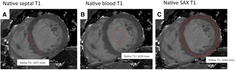
Healthy subjects (n=102; mean age 41 years (range 17-83), male, n=53 (52%)), with no previous medical history, and normotensive low risk subjects (n=113) referred for clinical cardiovascular magnetic resonance (CMR) were examined. Further inclusion criteria for all were absence of regular medication and subsequently normal findings of routine CMR. All subjects underwent T1 mapping using a uniform imaging set-up (modified Look- Locker inversion recovery, MOLLI, using scheme 3(3)3(3)5)) on 1.5 Tesla (T) and 3 T Philips scanners. Native T1-maps were acquired in a single midventricular short axis slice and repeated 20 minutes following gadobutrol. Reference values were obtained for native T1 and gadolinium-based partition coefficients, λ and extracellular volume fraction (ECV) in a core lab using standardized postprocessing.

RESULTS

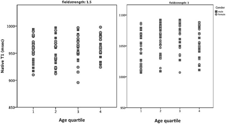
In healthy controls, mean native T1 values were 950±21 msec at 1.5 T and 1052±23 at 3 T. λ and ECV values were 0.44±0.06 and 0.25±0.04 at 1.5 T, and 0.44±0.07 and 0.26±0.04 at 3 T, respectively. There were no significant differences between healthy controls and low risk subjects in routine CMR parameters and T1 values. The entire cohort showed no correlation between age, gender and native T1. Cross-center comparisons of mean values showed no significant difference for any of the T1 indices at any field strength. There were considerable regional differences in segmental T1 values. λ and ECV were found to be dose dependent. There was excellent inter- and intraobserver reproducibility for measurement of native septal T1.

CONCLUSION

We show transferability for a unifying T1 mapping methodology in a multicenter setting. We provide reference ranges for T1 values in healthy human myocardium, which can be applied across participating sites.

摘要

背景

T1 映射是一种强大且高度可重复的应用,用于量化心肌纵向磁化的弛豫。目前可用的 T1 映射方法因设备和供应商而异,不同系统和序列之间 T1 值的准确性和精密度各不相同。我们评估了一种 T1 映射方法的可转移性,并在多中心环境中确定了健康人心肌的参考值。

方法

对健康受试者(n = 102;平均年龄 41 岁(范围 17 - 83 岁),男性,n = 53(52%))进行检查,这些受试者无既往病史,以及因临床心血管磁共振(CMR)检查而转诊的血压正常的低风险受试者(n = 113)。所有受试者的进一步纳入标准为未规律用药且随后常规 CMR 检查结果正常。所有受试者在 1.5 特斯拉(T)和 3T 的飞利浦扫描仪上使用统一的成像设置(改良 Look - Locker 反转恢复序列,MOLLI,采用 3(3)3(3)5 方案)进行 T1 映射。在单个心室中部短轴切片中获取原始 T1 图,并在注射钆布醇后 20 分钟重复采集。在核心实验室使用标准化后处理获得原始 T1、基于钆的分配系数λ和细胞外体积分数(ECV)的参考值。

结果

在健康对照组中,1.5T 时平均原始 T1 值为 950±21 毫秒,3T 时为 1052±23 毫秒。1.5T 时λ和 ECV 值分别为 0.44±0.06 和 0.25±0.04,3T 时分别为 0.44±0.07 和 0.26±0.04。健康对照组和低风险受试者在常规 CMR 参数和 T1 值方面无显著差异。整个队列中年龄、性别与原始 T1 之间无相关性。平均值的跨中心比较显示在任何场强下任何 T1 指标均无显著差异。节段性 T1 值存在相当大的区域差异。发现λ和 ECV 与剂量相关。原始间隔 T1 测量的观察者间和观察者内再现性都非常好。

结论

我们展示了一种统一的 T1 映射方法在多中心环境中的可转移性。我们提供了健康人心肌 T1 值的参考范围,可应用于各参与站点。

https://cdn.ncbi.nlm.nih.gov/pmc/blobs/8100/4203908/50fc98b262f0/12968_2014_69_Fig7_HTML.jpg
https://cdn.ncbi.nlm.nih.gov/pmc/blobs/8100/4203908/e61e11059fa5/12968_2014_69_Fig1_HTML.jpg
https://cdn.ncbi.nlm.nih.gov/pmc/blobs/8100/4203908/2af636d1d332/12968_2014_69_Fig2_HTML.jpg
https://cdn.ncbi.nlm.nih.gov/pmc/blobs/8100/4203908/a435afca1104/12968_2014_69_Fig3_HTML.jpg
https://cdn.ncbi.nlm.nih.gov/pmc/blobs/8100/4203908/ae680df4f005/12968_2014_69_Fig4_HTML.jpg
https://cdn.ncbi.nlm.nih.gov/pmc/blobs/8100/4203908/5e5c2656a36a/12968_2014_69_Fig5_HTML.jpg
https://cdn.ncbi.nlm.nih.gov/pmc/blobs/8100/4203908/1243462856d6/12968_2014_69_Fig6_HTML.jpg
https://cdn.ncbi.nlm.nih.gov/pmc/blobs/8100/4203908/50fc98b262f0/12968_2014_69_Fig7_HTML.jpg

相似文献

1
Reference values for healthy human myocardium using a T1 mapping methodology: results from the International T1 Multicenter cardiovascular magnetic resonance study.使用T1映射方法测定健康人心肌的参考值:国际T1多中心心血管磁共振研究结果
J Cardiovasc Magn Reson. 2014 Oct 21;16(1):69. doi: 10.1186/s12968-014-0069-x.
2
Myocardial T-mapping at 3T using saturation-recovery: reference values, precision and comparison with MOLLI.使用饱和恢复法在3T下进行心肌T值映射:参考值、精密度及与MOLLI的比较
J Cardiovasc Magn Reson. 2016 Nov 18;18(1):84. doi: 10.1186/s12968-016-0302-x.
3
Z-score mapping for standardized analysis and reporting of cardiovascular magnetic resonance modified Look-Locker inversion recovery (MOLLI) T1 data: Normal behavior and validation in patients with amyloidosis.Z 分数映射用于心血管磁共振改良 Look-Locker 反转恢复(MOLLI)T1 数据的标准化分析和报告:淀粉样变性患者的正常表现和验证。
J Cardiovasc Magn Reson. 2020 Jan 20;22(1):6. doi: 10.1186/s12968-019-0595-7.
4
Normal variation of magnetic resonance T1 relaxation times in the human population at 1.5 T using ShMOLLI.使用 ShMOLLI 技术在 1.5T 场强下人脑中磁共振 T1 弛豫时间的正常变异。
J Cardiovasc Magn Reson. 2013 Jan 20;15(1):13. doi: 10.1186/1532-429X-15-13.
5
T-Mapping and extracellular volume estimates in pediatric subjects with Duchenne muscular dystrophy and healthy controls at 3T.3T 下杜氏肌营养不良症患儿与健康对照者的 T 映射和细胞外容积评估。
J Cardiovasc Magn Reson. 2020 Dec 10;22(1):85. doi: 10.1186/s12968-020-00687-z.
6
Standardization of T1 measurements with MOLLI in differentiation between health and disease--the ConSept study.MOLLI 测量 T1 值在健康与疾病鉴别诊断中的标准化——ConSept 研究。
J Cardiovasc Magn Reson. 2013 Sep 11;15(1):78. doi: 10.1186/1532-429X-15-78.
7
T1 mapping of the myocardium: intra-individual assessment of post-contrast T1 time evolution and extracellular volume fraction at 3T for Gd-DTPA and Gd-BOPTA.心肌 T1 映射:3T 下钆-DTPA 和钆-BOPTA 对比后 T1 时间演变和细胞外容积分数的个体内评估。
J Cardiovasc Magn Reson. 2012 Apr 28;14(1):26. doi: 10.1186/1532-429X-14-26.
8
Real-time cardiovascular magnetic resonance T1 and extracellular volume fraction mapping for tissue characterisation in aortic stenosis.实时心血管磁共振T1和细胞外容积分数成像用于主动脉瓣狭窄的组织特征分析
J Cardiovasc Magn Reson. 2020 Jun 22;22(1):46. doi: 10.1186/s12968-020-00632-0.
9
Inter-scanner comparability of Z-scores for native myocardial T1 and T2 mapping.不同扫描仪间 native 心肌 T1 和 T2 映射的 Z 评分的可重复性。
J Cardiovasc Magn Reson. 2024 Summer;26(1):100004. doi: 10.1016/j.jocmr.2023.100004. Epub 2024 Jan 9.
10
Comparison of different cardiovascular magnetic resonance sequences for native myocardial T1 mapping at 3T.3T下用于天然心肌T1映射的不同心血管磁共振序列的比较
J Cardiovasc Magn Reson. 2016 Oct 4;18(1):65. doi: 10.1186/s12968-016-0286-6.

引用本文的文献

1
Implications of Myocardial Fibrosis Burden on Left Ventricular Systolic Function in Sepsis Survivors: Insights from a Retrospective Cohort Study Using Quantitative Late Gadolinium Enhancement Cardiovascular Magnetic Resonance.脓毒症幸存者中心肌纤维化负担对左心室收缩功能的影响:一项使用定量延迟钆增强心血管磁共振的回顾性队列研究的见解
J Cardiovasc Dev Dis. 2025 Aug 13;12(8):306. doi: 10.3390/jcdd12080306.
2
Diagnostic performance of cardiovascular magnetic resonance parametric mapping as per modified Lake Louise Criteria in acute myocarditis: an updated systematic review and meta-analysis.根据改良的路易斯湖标准,心血管磁共振参数成像在急性心肌炎中的诊断性能:一项更新的系统评价和荟萃分析。
J Cardiovasc Imaging. 2025 Jun 3;33(1):5. doi: 10.1186/s44348-025-00048-3.
3

本文引用的文献

1
Aortic stiffness and interstitial myocardial fibrosis by native T1 are independently associated with left ventricular remodeling in patients with dilated cardiomyopathy.在扩张型心肌病患者中,主动脉僵硬度和基于固有T1的心肌间质纤维化与左心室重构独立相关。
Hypertension. 2014 Oct;64(4):762-8. doi: 10.1161/HYPERTENSIONAHA.114.03928. Epub 2014 Jul 14.
2
T1 mapping - beware regional variations.T1 映射——注意区域差异。
Eur Heart J Cardiovasc Imaging. 2014 Nov;15(11):1302. doi: 10.1093/ehjci/jeu082. Epub 2014 May 22.
3
Individualized cardiovascular risk assessment by cardiovascular magnetic resonance.
Cardiac magnetic resonance in heart transplant recipients: histological, clinical and cell-free DNA validation.心脏移植受者的心脏磁共振成像:组织学、临床及游离DNA验证
Eur Heart J Cardiovasc Imaging. 2025 Jul 31;26(8):1420-1428. doi: 10.1093/ehjci/jeaf145.
4
Myocardial native T1 and extracellular volume measurements at 5T: Feasibility study and initial experience.5T场强下心肌固有T1和细胞外容积测量:可行性研究与初步经验
J Cardiovasc Magn Reson. 2025;27(1):101896. doi: 10.1016/j.jocmr.2025.101896. Epub 2025 Apr 21.
5
Native T1 mapping times are strongly influenced by elite athlete status.固有T1映射时间受精英运动员身份的强烈影响。
Eur Heart J Cardiovasc Imaging. 2025 Jun 30;26(7):1208-1216. doi: 10.1093/ehjci/jeaf123.
6
Liver T1 mapping in Fontan patients and patients with biventricular congenital heart disease - insights into the effects of venous congestions on diffuse liver disease.Fontan患者和双心室先天性心脏病患者的肝脏T1 mapping——关于静脉淤血对弥漫性肝病影响的见解
Int J Cardiovasc Imaging. 2025 Feb;41(2):347-358. doi: 10.1007/s10554-024-03314-5. Epub 2025 Jan 8.
7
Assessment of Myocardial Fibrosis in Marfan Syndrome Using Cardiac Magnetic Resonance Imaging.应用心脏磁共振成像评估马凡综合征患者的心肌纤维化。
Mol Genet Genomic Med. 2024 Nov;12(11):e70024. doi: 10.1002/mgg3.70024.
8
NT-proBNP Reflects Left Ventricular Hypertrophy Rather than Left Ventricular Dilatation or Systolic Dysfunction in Patients with Fabry Disease.N末端B型利钠肽原反映法布里病患者的左心室肥厚,而非左心室扩张或收缩功能障碍。
J Clin Med. 2024 Oct 7;13(19):5953. doi: 10.3390/jcm13195953.
9
Basal inferoseptal segment is highly susceptible to deformation in the clinical spectrum of transthyretin-derived amyloid cardiomyopathy.在转甲状腺素蛋白源性淀粉样心肌病的临床谱系中,基底室间隔下壁节段极易发生变形。
Eur Heart J Open. 2024 Sep 2;4(5):oeae076. doi: 10.1093/ehjopen/oeae076. eCollection 2024 Sep.
10
Analysis of myocardial T1, T2, and T2* values by age, sex, and cardiac segments in normal population: a prospective study.正常人群中心肌 T1、T2 和 T2* 值的分析:一项前瞻性研究。
Int J Cardiovasc Imaging. 2024 Nov;40(11):2357-2369. doi: 10.1007/s10554-024-03241-5. Epub 2024 Sep 16.
通过心血管磁共振进行个体化心血管风险评估。
Future Cardiol. 2014 Mar;10(2):273-89. doi: 10.2217/fca.13.102.
4
T1-mapping in the heart: accuracy and precision.心脏 T1-mapping:准确性和精密度。
J Cardiovasc Magn Reson. 2014 Jan 4;16(1):2. doi: 10.1186/1532-429X-16-2.
5
Myocardial T1 mapping and extracellular volume quantification: a Society for Cardiovascular Magnetic Resonance (SCMR) and CMR Working Group of the European Society of Cardiology consensus statement.心肌 T1 映射和细胞外容积定量:心血管磁共振学会 (SCMR) 和欧洲心脏病学会 CMR 工作组的共识声明。
J Cardiovasc Magn Reson. 2013 Oct 14;15(1):92. doi: 10.1186/1532-429X-15-92.
6
Cardiac magnetic resonance postcontrast T1 time is associated with outcome in patients with heart failure and preserved ejection fraction.心脏磁共振钆对比增强 T1 时间与射血分数保留心力衰竭患者的结局相关。
Circ Cardiovasc Imaging. 2013 Nov;6(6):1056-65. doi: 10.1161/CIRCIMAGING.113.000633. Epub 2013 Sep 13.
7
Standardization of T1 measurements with MOLLI in differentiation between health and disease--the ConSept study.MOLLI 测量 T1 值在健康与疾病鉴别诊断中的标准化——ConSept 研究。
J Cardiovasc Magn Reson. 2013 Sep 11;15(1):78. doi: 10.1186/1532-429X-15-78.
8
CMR quantification of myocardial scar provides additive prognostic information in nonischemic cardiomyopathy.CMR 定量评估心肌瘢痕有助于提供非缺血性心肌病的预后信息。
JACC Cardiovasc Imaging. 2013 Sep;6(9):944-54. doi: 10.1016/j.jcmg.2013.05.013. Epub 2013 Aug 8.
9
Influence of Off-resonance in myocardial T1-mapping using SSFP based MOLLI method.基于 SSFP 技术的 MOLLI 方法测量心肌 T1 值时离频的影响。
J Cardiovasc Magn Reson. 2013 Jul 22;15(1):63. doi: 10.1186/1532-429X-15-63.
10
Evaluation of age-related interstitial myocardial fibrosis with cardiac magnetic resonance contrast-enhanced T1 mapping: MESA (Multi-Ethnic Study of Atherosclerosis).心脏磁共振对比增强 T1 映射评估年龄相关性间质心肌纤维化:MESA(动脉粥样硬化的多民族研究)。
J Am Coll Cardiol. 2013 Oct 1;62(14):1280-1287. doi: 10.1016/j.jacc.2013.05.078. Epub 2013 Jul 17.