• 文献检索
  • 文档翻译
  • 深度研究
  • 学术资讯
  • Suppr Zotero 插件Zotero 插件
  • 邀请有礼
  • 套餐&价格
  • 历史记录
应用&插件
Suppr Zotero 插件Zotero 插件浏览器插件Mac 客户端Windows 客户端微信小程序
定价
高级版会员购买积分包购买API积分包
服务
文献检索文档翻译深度研究API 文档MCP 服务
关于我们
关于 Suppr公司介绍联系我们用户协议隐私条款
关注我们

Suppr 超能文献

核心技术专利:CN118964589B侵权必究
粤ICP备2023148730 号-1Suppr @ 2026

文献检索

告别复杂PubMed语法,用中文像聊天一样搜索,搜遍4000万医学文献。AI智能推荐,让科研检索更轻松。

立即免费搜索

文件翻译

保留排版,准确专业,支持PDF/Word/PPT等文件格式,支持 12+语言互译。

免费翻译文档

深度研究

AI帮你快速写综述,25分钟生成高质量综述,智能提取关键信息,辅助科研写作。

立即免费体验

MOLLI 测量 T1 值在健康与疾病鉴别诊断中的标准化——ConSept 研究。

Standardization of T1 measurements with MOLLI in differentiation between health and disease--the ConSept study.

机构信息

Department of Cardiovascular Imaging, King's College London, London, UK.

出版信息

J Cardiovasc Magn Reson. 2013 Sep 11;15(1):78. doi: 10.1186/1532-429X-15-78.

DOI:10.1186/1532-429X-15-78
PMID:24025486
原文链接:https://pmc.ncbi.nlm.nih.gov/articles/PMC3847466/
Abstract

BACKGROUND

T1 imaging based on pixel-wise quantification of longitudinal relaxation has the potential to differentiate between normal and abnormal myocardium. The accuracy of T1 measurement has not been established nor systematically tested in the presence of health and disease.

METHODS

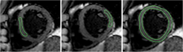
Intra-observer, inter-observer and inter-study reproducibility of T1 imaging was assessed in subjects with left ventricular hypertrophy (LVH, n = 25) or dilated cardiomyopathy (DCM, n = 43). Thirty-eight subjects with low-pretest likelihood of cardiomyopathy served as a control group. T1 values were acquired in a single mid-ventricular short axis slice using modified Look-Locker imaging prior and after the application of gadolinium contrast at 1.5 and 3 T. Analysis was performed with regions of interest (ROI) placed conservatively within the septum or to include the whole short axis (SAX) myocardium.

RESULTS

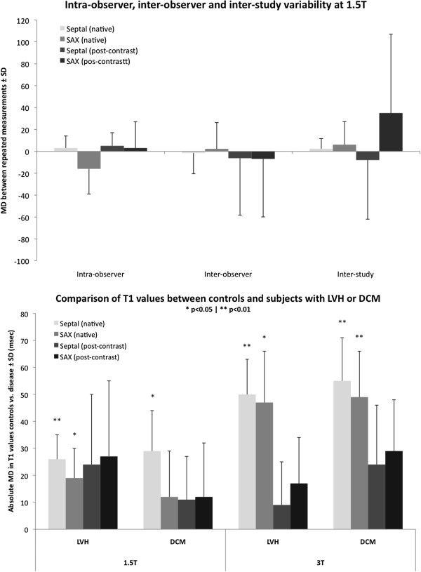
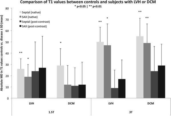
Intra-observer, inter-observer and inter-study repeated measurements within the septum showed smaller mean differences and narrower 95% confidence intervals than repeated short axis ROI measurements. Native T1 values were higher in septal ROIs compared with SAX values at both field strengths (1.5 T: 976 ± 37 vs. 952 ± 41, p < 0.01; 3 T: 1108 ± 67 vs. 1087 ± 60, p < 0.01). Native T1 values revealed significant mean differences between controls and patients with LVH for both septal (1.5 T: 26 ± 9, p < 0.01; 3 T: 50 ± 13, p < 0.01) and SAX ROIs (1.5 T: 19 ± 11, p < 0.05; 3 T: 47 ± 19, p < 0.05) with greater differences observed at 3 T versus 1.5 T field strength. Native T1 values revealed significant mean differences between controls and patients with DCM for septal ROI (1.5 T: 29 ± 15, p < 0.05; 3 T: 55 ± 16, p < 0.01) at both 1.5 T and 3 T, but only for SAX ROIs at 3 T (49 ± 17, p < 0.01). There were no significant differences in post-contrast T1 values or partition coefficient (λ) between controls and patients.

CONCLUSION

Conservative septal ROI T1 measurement is a robust technique with excellent intra-observer, inter-observer and inter-study reproducibility for native and post-contrast T1 value and partition coefficient measurements. Moreover, native septal T1 values reveal the greatest difference between normal and abnormal myocardium, which is independent of geometrical alterations of cardiac chamber and wall thickness. We propose the use of native T1 measurements using conservative septal technique as the standardized approach to distinguish health from disease assuming diffuse myocardial involvement.

摘要

背景

基于像素的纵向弛豫定量的 T1 成像有可能区分正常和异常心肌。T1 测量的准确性尚未在健康和疾病存在的情况下得到确定或系统测试。

方法

在左心室肥厚 (LVH,n = 25) 或扩张型心肌病 (DCM,n = 43) 患者中评估 T1 成像的观察者内、观察者间和研究间可重复性。38 名低心肌病变可能性的受试者作为对照组。使用改良的 Look-Locker 成像技术,在 1.5 和 3 T 时,在单个中室短轴切片上获取 T1 值,并在应用钆对比剂前后进行。使用感兴趣区域 (ROI) 进行分析,ROI 放置在间隔内保守,或包括整个短轴 (SAX) 心肌。

结果

间隔内的观察者内、观察者间和研究间重复测量的平均差异较小,95%置信区间较狭窄,而 SAX ROI 测量的重复测量的平均差异较大。在两种场强下,间隔 ROI 的 T1 值均高于 SAX 值 (1.5 T:976 ± 37 与 952 ± 41,p < 0.01;3 T:1108 ± 67 与 1087 ± 60,p < 0.01)。在两种场强下,与对照组相比,LVH 患者的 T1 值均有显著的平均差异,无论是间隔 (1.5 T:26 ± 9,p < 0.01;3 T:50 ± 13,p < 0.01) 还是 SAX ROI (1.5 T:19 ± 11,p < 0.05;3 T:47 ± 19,p < 0.05),在 3 T 时差异大于 1.5 T。在两种场强下,与对照组相比,DCM 患者的 T1 值均有显著的平均差异,间隔 ROI 有差异 (1.5 T:29 ± 15,p < 0.05;3 T:55 ± 16,p < 0.01),而在 3 T 时 SAX ROI 有差异 (49 ± 17,p < 0.01)。在对照组和患者之间,没有观察到钆后 T1 值或分割系数 (λ) 的差异。

结论

采用保守的间隔 ROI T1 测量技术,对于 T1 值和分割系数的固有和钆后测量,具有出色的观察者内、观察者间和研究间可重复性。此外,间隔的固有 T1 值显示了正常和异常心肌之间的最大差异,这与心脏腔室和壁厚度的几何变化无关。我们建议使用保守的间隔技术测量固有 T1 值,作为区分健康与疾病的标准方法,假设弥漫性心肌受累。

https://cdn.ncbi.nlm.nih.gov/pmc/blobs/bbed/3847466/84342c90b966/1532-429X-15-78-3.jpg
https://cdn.ncbi.nlm.nih.gov/pmc/blobs/bbed/3847466/d6cb2de3abfd/1532-429X-15-78-1.jpg
https://cdn.ncbi.nlm.nih.gov/pmc/blobs/bbed/3847466/b12d36b271c9/1532-429X-15-78-2.jpg
https://cdn.ncbi.nlm.nih.gov/pmc/blobs/bbed/3847466/84342c90b966/1532-429X-15-78-3.jpg
https://cdn.ncbi.nlm.nih.gov/pmc/blobs/bbed/3847466/d6cb2de3abfd/1532-429X-15-78-1.jpg
https://cdn.ncbi.nlm.nih.gov/pmc/blobs/bbed/3847466/b12d36b271c9/1532-429X-15-78-2.jpg
https://cdn.ncbi.nlm.nih.gov/pmc/blobs/bbed/3847466/84342c90b966/1532-429X-15-78-3.jpg

相似文献

1
Standardization of T1 measurements with MOLLI in differentiation between health and disease--the ConSept study.MOLLI 测量 T1 值在健康与疾病鉴别诊断中的标准化——ConSept 研究。
J Cardiovasc Magn Reson. 2013 Sep 11;15(1):78. doi: 10.1186/1532-429X-15-78.
2
Comparative study on native T value of myocardium between using ConSept and SAX approaches in non-ischemic dilated cardiomyopathy.非缺血性扩张型心肌病中 ConSept 和 SAX 方法测量心肌固有 T 值的对比研究。
Zhong Nan Da Xue Xue Bao Yi Xue Ban. 2021;46(9):1003-1009. doi: 10.11817/j.issn.1672-7347.2021.210229.
3
Native T1 mapping in differentiation of normal myocardium from diffuse disease in hypertrophic and dilated cardiomyopathy.心肌肥厚型和扩张型心肌病中正常心肌与弥漫性病变的 T1 自然映射鉴别。
JACC Cardiovasc Imaging. 2013 Apr;6(4):475-84. doi: 10.1016/j.jcmg.2012.08.019. Epub 2013 Mar 14.
4
Loss of base-to-apex circumferential strain gradient assessed by cardiovascular magnetic resonance in Fabry disease: relationship to T1 mapping, late gadolinium enhancement and hypertrophy.通过心血管磁共振评估法布里病的基底-顶点周向应变梯度缺失:与 T1 映射、晚期钆增强和心肌肥厚的关系。
J Cardiovasc Magn Reson. 2019 Aug 1;21(1):45. doi: 10.1186/s12968-019-0557-0.
5
Reference values for healthy human myocardium using a T1 mapping methodology: results from the International T1 Multicenter cardiovascular magnetic resonance study.使用T1映射方法测定健康人心肌的参考值:国际T1多中心心血管磁共振研究结果
J Cardiovasc Magn Reson. 2014 Oct 21;16(1):69. doi: 10.1186/s12968-014-0069-x.
6
Z-score mapping for standardized analysis and reporting of cardiovascular magnetic resonance modified Look-Locker inversion recovery (MOLLI) T1 data: Normal behavior and validation in patients with amyloidosis.Z 分数映射用于心血管磁共振改良 Look-Locker 反转恢复(MOLLI)T1 数据的标准化分析和报告:淀粉样变性患者的正常表现和验证。
J Cardiovasc Magn Reson. 2020 Jan 20;22(1):6. doi: 10.1186/s12968-019-0595-7.
7
T1 mapping of the myocardium: intra-individual assessment of the effect of field strength, cardiac cycle and variation by myocardial region.心肌 T1 映射:个体内评估场强、心动周期和心肌区域变化的影响。
J Cardiovasc Magn Reson. 2012 May 1;14(1):27. doi: 10.1186/1532-429X-14-27.
8
Myocardial T-mapping at 3T using saturation-recovery: reference values, precision and comparison with MOLLI.使用饱和恢复法在3T下进行心肌T值映射:参考值、精密度及与MOLLI的比较
J Cardiovasc Magn Reson. 2016 Nov 18;18(1):84. doi: 10.1186/s12968-016-0302-x.
9
Towards standardized postprocessing of global longitudinal strain by feature tracking - OptiStrain CMR-FT study.面向特征追踪的全球纵向应变标准化后处理 - OptiStrain CMR-FT 研究。
BMC Cardiovasc Disord. 2019 Nov 27;19(1):267. doi: 10.1186/s12872-019-1255-4.
10
SASHA versus ShMOLLI: a comparison of T1 mapping methods in health and dilated cardiomyopathy at 3 T.SASHA与ShMOLLI对比:3T下健康及扩张型心肌病中T1映射方法的比较
Int J Cardiovasc Imaging. 2017 Oct;33(10):1551-1560. doi: 10.1007/s10554-017-1134-y. Epub 2017 Jun 6.

引用本文的文献

1
Clinical utility of segmental native T1 mapping for diagnosis and risk-stratification in cardiac sarcoidosis.节段性心肌T1值测绘在心脏结节病诊断及风险分层中的临床应用价值
Int J Cardiol Heart Vasc. 2025 Aug 29;61:101787. doi: 10.1016/j.ijcha.2025.101787. eCollection 2025 Dec.
2
Native T1 adds independent value for cardiovascular risk assessment beyond global longitudinal strain in an all-comers real-world clinical patient population.在所有患者的真实临床患者群体中,相较于整体纵向应变,固有T1为心血管风险评估增加了独立价值。
Eur Heart J Open. 2025 Aug 20;5(5):oeaf109. doi: 10.1093/ehjopen/oeaf109. eCollection 2025 Sep.
3

本文引用的文献

1
Native T1 mapping in differentiation of normal myocardium from diffuse disease in hypertrophic and dilated cardiomyopathy.心肌肥厚型和扩张型心肌病中正常心肌与弥漫性病变的 T1 自然映射鉴别。
JACC Cardiovasc Imaging. 2013 Apr;6(4):475-84. doi: 10.1016/j.jcmg.2012.08.019. Epub 2013 Mar 14.
2
Noncontrast T1 mapping for the diagnosis of cardiac amyloidosis.非对比 T1 映射在心脏淀粉样变性诊断中的应用。
JACC Cardiovasc Imaging. 2013 Apr;6(4):488-97. doi: 10.1016/j.jcmg.2012.11.013. Epub 2013 Mar 14.
3
Native myocardial T1 mapping by cardiovascular magnetic resonance imaging in subclinical cardiomyopathy in patients with systemic lupus erythematosus.
Serial cardiac magnetic resonance imaging to assess myocardial inflammation in PET-positive cardiac sarcoidosis under immunomodulatory therapy: a retrospective observational study.
采用连续心脏磁共振成像评估免疫调节治疗下PET阳性心脏结节病的心肌炎症:一项回顾性观察研究。
BMJ Open. 2025 Aug 25;15(8):e098540. doi: 10.1136/bmjopen-2024-098540.
4
Diagnostic usefulness and limitation of cardiac magnetic resonance for identifying myocardial damage in survivors of cardiac arrest in midtown.心脏磁共振成像在识别市中心心脏骤停幸存者心肌损伤方面的诊断价值与局限性
World J Radiol. 2025 Jun 28;17(6):104473. doi: 10.4329/wjr.v17.i6.104473.
5
Neural networks with personalized training for improved MOLLI T mapping.具有个性化训练以改进MOLLI T映射的神经网络。
BMC Med Imaging. 2025 Jul 1;25(1):245. doi: 10.1186/s12880-025-01769-z.
6
Advancing Myocardial T1 Mapping: A Comparative Study of the Frequency-Independent MFA Sequence and Standard MOLLI.心肌T1映射技术的进展:频率无关MFA序列与标准MOLLI的比较研究
NMR Biomed. 2025 May;38(5):e70031. doi: 10.1002/nbm.70031.
7
3D joint T/T /T mapping and water-fat imaging for contrast-agent free myocardial tissue characterization at 1.5T.1.5T下用于无对比剂心肌组织特征分析的三维关节T/T/T映射及水脂成像
Magn Reson Med. 2025 Jun;93(6):2297-2310. doi: 10.1002/mrm.30397. Epub 2025 Feb 21.
8
The Protocol for the Multi-Ethnic, multi-centre raNdomised controlled trial of a low-energy Diet for improving functional status in heart failure with Preserved ejection fraction (AMEND Preserved).多民族、多中心随机对照试验方案:低能量饮食改善射血分数保留的心力衰竭患者功能状态(AMEND保留方案)
BMJ Open. 2025 Jan 28;15(1):e094722. doi: 10.1136/bmjopen-2024-094722.
9
Elevated septal native T1 time in cardiac magnetic resonance imaging suggesting myocardial fibrosis in young kidney transplant recipients.心脏磁共振成像中室间隔固有T1时间升高提示年轻肾移植受者存在心肌纤维化。
J Cardiovasc Magn Reson. 2025 Jan 13;27(1):101839. doi: 10.1016/j.jocmr.2025.101839.
10
Design and rationale of MYOFLAME-19 randomised controlled trial: MYOcardial protection to reduce post-COVID inFLAMmatory heart disease using cardiovascular magnetic resonance Endpoints.MYOFLAME-19随机对照试验的设计与原理:利用心血管磁共振终点指标进行心肌保护以减少新冠后炎症性心脏病
J Cardiovasc Magn Reson. 2024 Oct 29;27(1):101121. doi: 10.1016/j.jocmr.2024.101121.
心血管磁共振成像技术在系统性红斑狼疮亚临床心肌病患者中心肌 T1 mapping 的应用。
Circ Cardiovasc Imaging. 2013 Mar 1;6(2):295-301. doi: 10.1161/CIRCIMAGING.112.000151. Epub 2013 Feb 12.
4
Diffuse myocardial fibrosis evaluation using cardiac magnetic resonance T1 mapping: sample size considerations for clinical trials.使用心脏磁共振 T1 映射评估弥漫性心肌纤维化:临床试验的样本量考虑。
J Cardiovasc Magn Reson. 2012 Dec 28;14(1):90. doi: 10.1186/1532-429X-14-90.
5
Myocardial tissue characterization using magnetic resonance noncontrast t1 mapping in hypertrophic and dilated cardiomyopathy.应用心脏磁共振钆对比增强 T1 mapping 技术对肥厚型和扩张型心肌病进行心肌组织特征分析。
Circ Cardiovasc Imaging. 2012 Nov;5(6):726-33. doi: 10.1161/CIRCIMAGING.112.976738. Epub 2012 Oct 15.
6
Extracellular volume fraction mapping in the myocardium, part 2: initial clinical experience.心肌细胞外容积分数测绘 2 部分:初步临床经验。
J Cardiovasc Magn Reson. 2012 Sep 11;14(1):64. doi: 10.1186/1532-429X-14-64.
7
Left ventricular chamber dimensions and wall thickness by cardiovascular magnetic resonance: comparison with transthoracic echocardiography.心血管磁共振左心室腔尺寸和室壁厚度:与经胸超声心动图的比较。
Eur Heart J Cardiovasc Imaging. 2013 Mar;14(3):240-6. doi: 10.1093/ehjci/jes145. Epub 2012 Jul 19.
8
T1 mapping of the myocardium: intra-individual assessment of the effect of field strength, cardiac cycle and variation by myocardial region.心肌 T1 映射:个体内评估场强、心动周期和心肌区域变化的影响。
J Cardiovasc Magn Reson. 2012 May 1;14(1):27. doi: 10.1186/1532-429X-14-27.
9
T1 mapping of the myocardium: intra-individual assessment of post-contrast T1 time evolution and extracellular volume fraction at 3T for Gd-DTPA and Gd-BOPTA.心肌 T1 映射:3T 下钆-DTPA 和钆-BOPTA 对比后 T1 时间演变和细胞外容积分数的个体内评估。
J Cardiovasc Magn Reson. 2012 Apr 28;14(1):26. doi: 10.1186/1532-429X-14-26.
10
Extracellular volume imaging by magnetic resonance imaging provides insights into overt and sub-clinical myocardial pathology.磁共振成像的细胞外容积成像是洞察显性和亚临床心肌病理学的一种手段。
Eur Heart J. 2012 May;33(10):1268-78. doi: 10.1093/eurheartj/ehr481. Epub 2012 Jan 24.