Hayden Rebecca S, Fortin Jean-Philippe, Harwood Benjamin, Subramanian Balajikarthick, Quinn Kyle P, Georgakoudi Irene, Kopin Alan S, Kaplan David L
4 Colby St., Medford, MA 02155 (USA).
800 Washington Street, Box 7703, Boston, MA 02111 (USA).
Adv Funct Mater. 2014 Jan 29;24(4):472-479. doi: 10.1002/adfm.201302210.
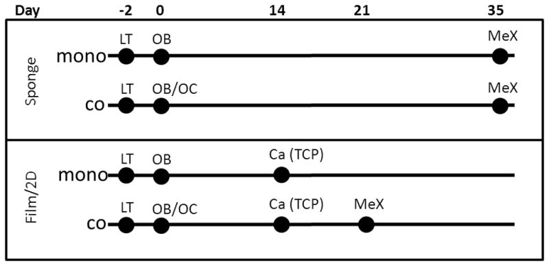
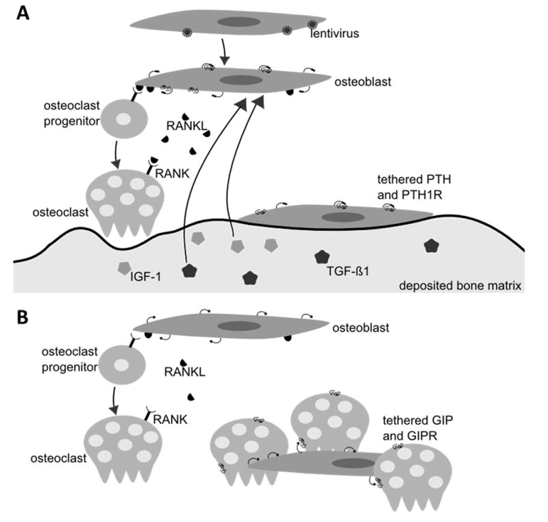
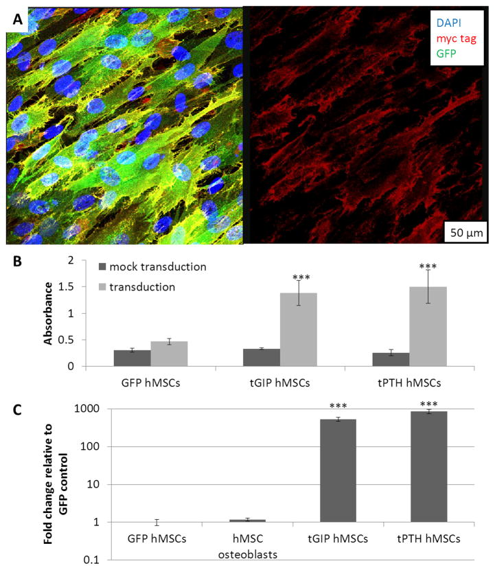
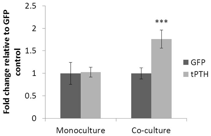
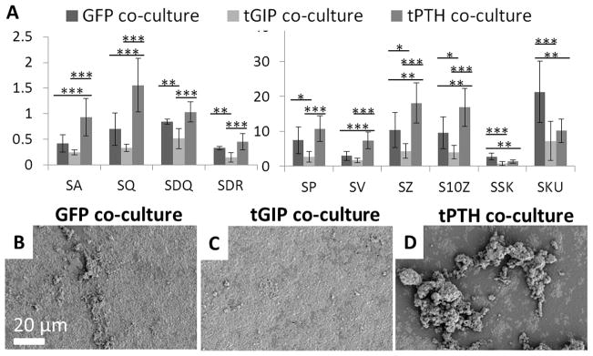
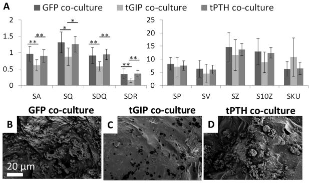
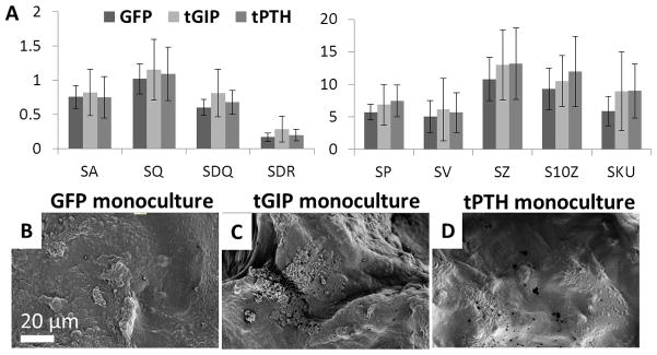
The goals of the present study are to establish an in vitro co-culture model of osteoblast and osteoclast function and to quantify the resulting bone remodeling. The bone is tissue engineered using well-defined silk protein biomaterials in 2D and 3D formats in combination with human cells expressing tethered agonists for selected G protein-coupled receptors (GPCRs). The tethered constructs are introduced with the objective of triggering sustained and localized GPCR signaling. The cell-modified biomaterial surfaces are reconstructed from SEM images into 3D models using image processing for quantitative measurement of surface characteristics. Parathyroid hormone (PTH) and glucose-dependent insulinotropic peptide (GIP) are selected because of their roles in bone remodeling for expression in tethered format on bone marrow derived human mesenchymal stem cells (hMSCs). Increased calcium deposition and increased surface roughness are found in 3D digital surface models constructed from SEM images of silk protein films remodeled by the co-cultures containing the tethered PTH, and decreased surface roughness is found for the films remodeled by the tethered GIP co-cultures. Increased surface roughness is not found in monocultures of hMSCs expressing tethered PTH, suggesting that osteoclast-osteoblast interactions in the presence of PTH signaling are responsible for the increased mineralization. These data point towards the design of in vitro bone models in which osteoblast-osteoclast interactions are mimicked for a better understanding of bone remodeling.
本研究的目标是建立成骨细胞和破骨细胞功能的体外共培养模型,并对由此产生的骨重塑进行量化。使用明确的丝蛋白生物材料以二维和三维形式与表达选定G蛋白偶联受体(GPCR)的拴系激动剂的人类细胞进行骨组织工程。引入拴系构建体的目的是触发持续和局部的GPCR信号传导。使用图像处理将细胞修饰的生物材料表面从扫描电子显微镜(SEM)图像重建为三维模型,以定量测量表面特征。选择甲状旁腺激素(PTH)和葡萄糖依赖性促胰岛素多肽(GIP),是因为它们在骨重塑中的作用,以便以拴系形式在人骨髓间充质干细胞(hMSC)上表达。在由含有拴系PTH的共培养物重塑的丝蛋白膜的SEM图像构建的三维数字表面模型中发现钙沉积增加和表面粗糙度增加,而在由拴系GIP共培养物重塑的膜中发现表面粗糙度降低。在表达拴系PTH的hMSC单培养物中未发现表面粗糙度增加,这表明在PTH信号存在下破骨细胞与成骨细胞的相互作用是矿化增加的原因。这些数据指向体外骨模型的设计,其中模拟破骨细胞与成骨细胞的相互作用以更好地理解骨重塑。