McElnea Elizabeth M, Hughes Emily, McGoldrick Aloysius, McCann Amanda, Quill Barry, Docherty Neil, Irnaten Mustapha, Farrell Michael, Clark Abbot F, O'Brien Colm J, Wallace Deborah M
University College Dublin School of Medicine and Health Sciences, University College Dublin, Dublin, Ireland.
BMC Ophthalmol. 2014 Dec 2;14:153. doi: 10.1186/1471-2415-14-153.
Disease associated alterations in the phenotype of lamina cribrosa (LC) cells are implicated in changes occurring at the optic nerve head (ONH) in glaucoma. Lipofuscin, the formation of which is driven by reactive oxygen species (ROS), is an intralysosomal, non-degradable, auto-fluorescent macromolecule which accumulates with age and can affect autophagy - the lysosomal degradation of a cell's constituents. We aimed to compare the content of lipofuscin-like material and markers of autophagy in LC cells from normal and glaucoma donor eyes.
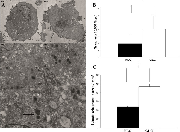
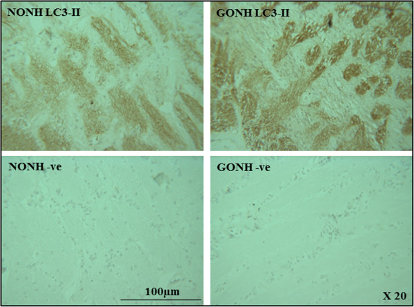
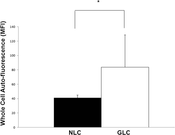
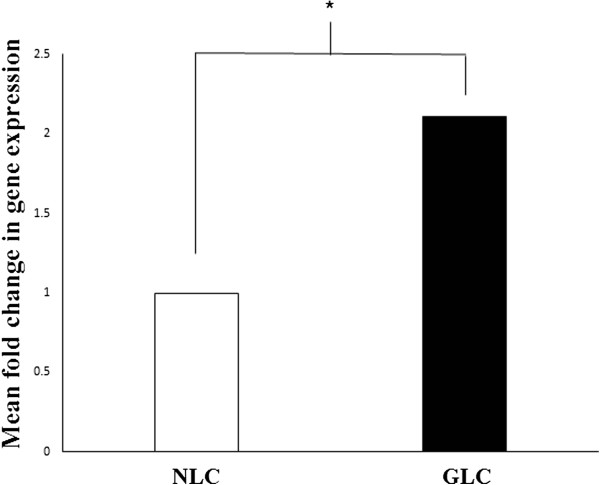
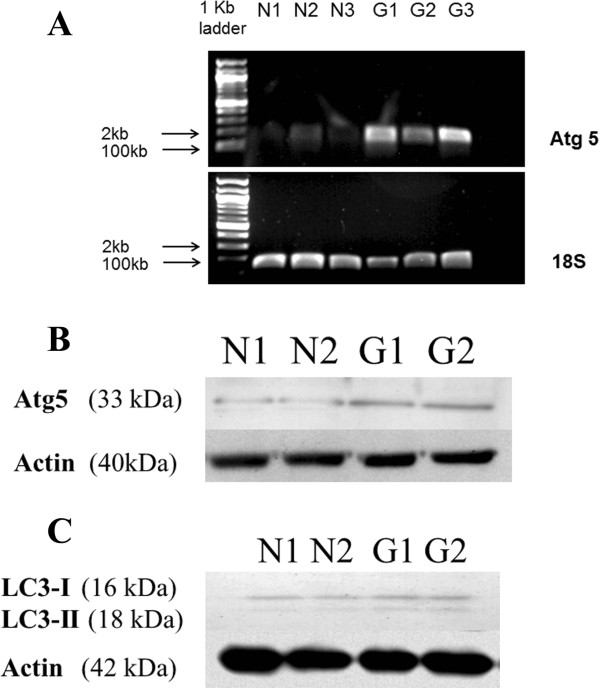
The number and size of peri-nuclear lysosomes were examined by transmission electron microscopy (TEM). Cellular auto-fluorescence was quantified by flow cytometry. Cathepsin K mRNA levels were assessed by PCR. Autophagy protein 5 (Atg5) mRNA and protein levels were analysed by PCR and Western blot. Protein levels of subunits of the microtubule associated proteins (MAP) 1A and 1B, light chain 3 (LC3) I and II were analysed by Western blot. Immunohistochemical staining of LC3-II in ONH sections from normal and glaucomatous donor eyes was performed.
A significant increase in the number of peri-nuclear lysosomes [4.1 × 10,000 per high power field (h.p.f.) ± 1.9 vs. 2.0 × 10,000 per h.p.f. ± 1.3, p = 0.002, n = 3] and whole cell auto-fluorescence (83.62 ± 45.1 v 41.01 ± 3.9, p = 0.02, n = 3) was found in glaucomatous LC cells relative to normal LC cells. Glaucomatous LC cells possessed significantly higher levels of Cathepsin K mRNA and Atg5 mRNA and protein. Enhanced levels of LC3-II were found in both LC cells and optic nerve head sections from glaucoma donors.
Increased lipofuscin formation is characteristic of LC cells from donors with glaucoma. This finding confirms the importance of oxidative stress in glaucoma pathogenesis. Intracellular lipofuscin accumulation may have important effects on autophagy the modification of which could form the basis for future novel glaucoma treatments.
筛板(LC)细胞表型的疾病相关改变与青光眼视神经乳头(ONH)发生的变化有关。脂褐素由活性氧(ROS)驱动形成,是一种溶酶体内的、不可降解的、自发荧光大分子,其会随年龄积累并可影响自噬——细胞成分的溶酶体降解。我们旨在比较正常和青光眼供体眼的LC细胞中脂褐素样物质的含量和自噬标志物。
通过透射电子显微镜(TEM)检查核周溶酶体的数量和大小。通过流式细胞术对细胞自发荧光进行定量。通过PCR评估组织蛋白酶K mRNA水平。通过PCR和蛋白质印迹分析自噬蛋白5(Atg5)的mRNA和蛋白质水平。通过蛋白质印迹分析微管相关蛋白(MAP)1A和1B亚基、轻链3(LC3)I和II的蛋白质水平。对正常和青光眼供体眼的ONH切片进行LC3-II的免疫组织化学染色。
与正常LC细胞相比,青光眼LC细胞中核周溶酶体数量显著增加[每高倍视野(h.p.f.)4.1×10,000±1.9 vs.每h.p.f. 2.0×10,000±1.3,p = 0.002,n = 3],全细胞自发荧光也显著增加(83.62±45.1对41.01±3.9,p = 0.02,n = 3)。青光眼LC细胞的组织蛋白酶K mRNA以及Atg5 mRNA和蛋白质水平显著更高。在青光眼供体的LC细胞和视神经乳头切片中均发现LC3-II水平升高。
脂褐素形成增加是青光眼供体LC细胞的特征。这一发现证实了氧化应激在青光眼发病机制中的重要性。细胞内脂褐素积累可能对自噬有重要影响,对其进行调节可能为未来新型青光眼治疗奠定基础。