Prestipino Filippo, Nenna Antonio, Casacalenda Adele, Chello Massimo
Cardiac Surgery Unit, University Hospital Campus Bio-Medico of Rome, Via Álvaro del Portillo, 21 - 00128 Roma, Italy.
Cardiac Surgery Unit, University Hospital Campus Bio-Medico of Rome, Via Álvaro del Portillo, 21 - 00128 Roma, Italy.
Int J Surg Case Rep. 2014;5(12):906-8. doi: 10.1016/j.ijscr.2014.10.006. Epub 2014 Oct 17.
Cardiac perforation is a rare, but potentially serious, complication of pacemaker implantation that may develop days or weeks after implantation.
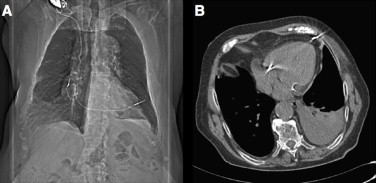
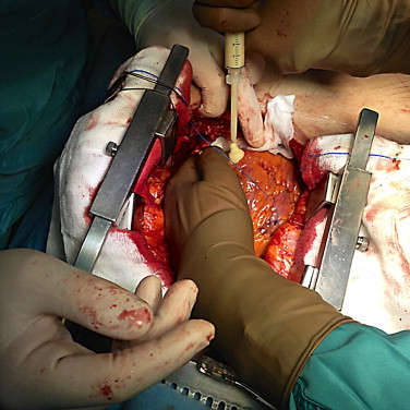
In the current case, 92-year-old man underwent permanent pacemaker implantation, but he presented 3 weeks later with severe symptoms. Computed tomography showed protrusion of the tip of the ventricular electrode through the right ventricle and into the chest wall. During an urgent surgical intervention, the lead was disconnected and extracted. A sealing hemostatic device and an hemostatic patch were applied to repair the ventricle; the procedure was uneventfull.
This case demonstrates how the correct diagnosis of ventricular perforation is crucial, and should be followed immediately by surgical planning.
The hemostatic patch is a valuable alternative to sutures in patients with thin and fragile ventricular wall, unable to undergo stitching.
心脏穿孔是起搏器植入术罕见但可能严重的并发症,可在植入后数天或数周出现。
在本病例中,一名92岁男性接受了永久性起搏器植入术,但3周后出现严重症状。计算机断层扫描显示心室电极尖端穿过右心室并突入胸壁。在紧急手术干预期间,导线被断开并取出。应用密封止血装置和止血贴片修复心室;手术过程顺利。
本病例表明正确诊断心室穿孔至关重要,随后应立即进行手术规划。
对于心室壁薄且脆弱、无法进行缝合的患者,止血贴片是缝合的一种有价值的替代方法。