Kim Dong-Yeon, Choi Yong-Seong, Kim Sung-Eun, Lee Jung-Ho, Kim Sue-Min, Kim Young-Jin, Rhie Jong-Won, Jun Young-Joon
Department of Plastic and Reconstructive Surgery, The Catholic University of Korea, Seoul, Korea.
Department of Orthopedic Surgery and Rare Diseases Institute, Korea University, Seoul, Korea.
J Korean Med Sci. 2014 Nov;29 Suppl 3(Suppl 3):S183-92. doi: 10.3346/jkms.2014.29.S3.S183. Epub 2014 Nov 21.
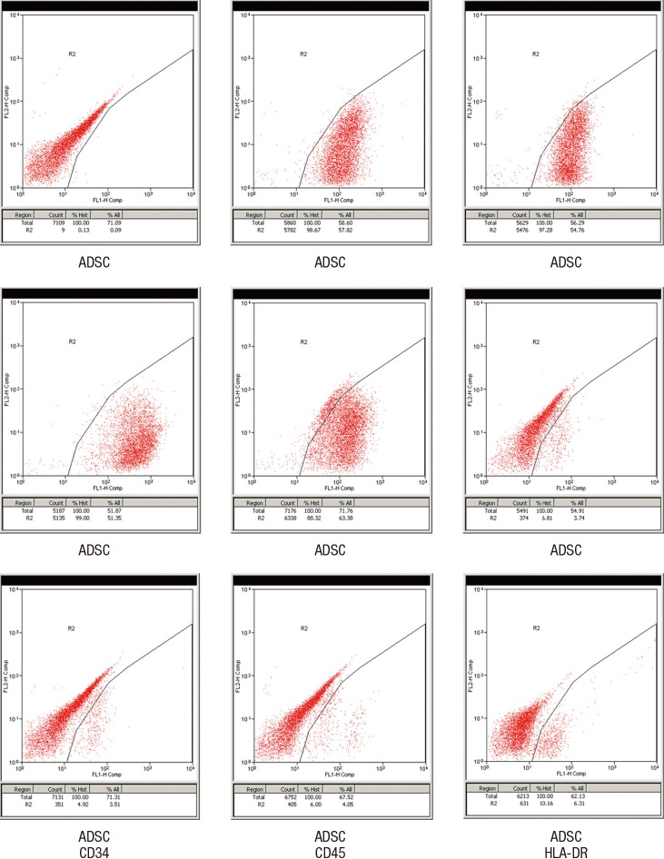
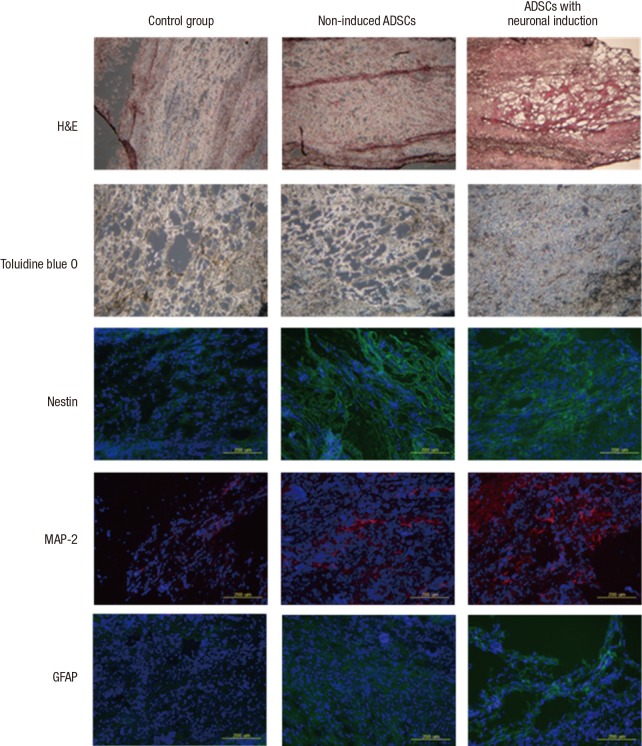
There have been many attempts for regeneration of peripheral nerve injury. In this study, we examined the in vivo effects of non-differentiated and neuronal differentiated adipose-derived stem cells (ADSCs) in inducing the neuronal regeneration in the Sprague-Dawley (SD) rats undergoing nerve defect bridged with the PCL nanotubes. Then, we performed immunohistochemical and histopathologic examinations, as well as the electromyography, in three groups: the control group (14 sciatic nerves transplanted with the PCL nanotube scaffold), the experimental group I (14 sciatic nerves with the non-differentiated ADSCs at a density of 7×10(5) cells/0.1 mL) and the experimental group II (14 sciatic nerves with the neuronal differentiated ADSCs at 7×10(5) cells/0.1 mL). Six weeks postoperatively, the degree of the neuronal induction and that of immunoreactivity to nestin, MAP-2 and GFAP was significantly higher in the experimental group I and II as compared with the control group. In addition, the nerve conduction velocity (NCV) was significantly higher in the experimental group I and II as compared with the control group (P=0.021 and P=0.020, respectively). On the other hand, there was no significant difference in the NCV between the two experimental groups (P>0.05). Thus, our results will contribute to treating patients with peripheral nerve defects using PCL nanotubes with ADSCs.
针对周围神经损伤的再生已经有许多尝试。在本研究中,我们检测了未分化和神经元分化的脂肪来源干细胞(ADSCs)在诱导接受聚己内酯(PCL)纳米管桥接神经缺损的Sprague-Dawley(SD)大鼠神经元再生中的体内效应。然后,我们对三组进行了免疫组织化学和组织病理学检查以及肌电图检查:对照组(14条坐骨神经移植了PCL纳米管支架)、实验组I(14条坐骨神经接种密度为7×10⁵ 个细胞/0.1 mL的未分化ADSCs)和实验组II(14条坐骨神经接种密度为7×10⁵ 个细胞/0.1 mL的神经元分化ADSCs)。术后六周,与对照组相比,实验组I和II中神经元诱导程度以及对巢蛋白、微管相关蛋白2(MAP-2)和胶质纤维酸性蛋白(GFAP)的免疫反应性程度显著更高。此外,与对照组相比,实验组I和II的神经传导速度(NCV)显著更高(分别为P = 0.021和P = 0.020)。另一方面,两个实验组之间的NCV没有显著差异(P>0.05)。因此,我们的结果将有助于使用PCL纳米管与ADSCs治疗周围神经缺损患者。