• 文献检索
  • 文档翻译
  • 深度研究
  • 学术资讯
  • Suppr Zotero 插件Zotero 插件
  • 邀请有礼
  • 套餐&价格
  • 历史记录
应用&插件
Suppr Zotero 插件Zotero 插件浏览器插件Mac 客户端Windows 客户端微信小程序
定价
高级版会员购买积分包购买API积分包
服务
文献检索文档翻译深度研究API 文档MCP 服务
关于我们
关于 Suppr公司介绍联系我们用户协议隐私条款
关注我们

Suppr 超能文献

核心技术专利:CN118964589B侵权必究
粤ICP备2023148730 号-1Suppr @ 2026

文献检索

告别复杂PubMed语法,用中文像聊天一样搜索,搜遍4000万医学文献。AI智能推荐,让科研检索更轻松。

立即免费搜索

文件翻译

保留排版,准确专业,支持PDF/Word/PPT等文件格式,支持 12+语言互译。

免费翻译文档

深度研究

AI帮你快速写综述,25分钟生成高质量综述,智能提取关键信息,辅助科研写作。

立即免费体验

使用水平集方法在二维超声心动图图像序列中测量左心室容积,以自动检测舒张末期和收缩末期帧。

Measuring Left Ventricular Volumes in Two-Dimensional Echocardiography Image Sequence Using Level-set Method for Automatic Detection of End-Diastole and End-systole Frames.

作者信息

Darvishi Saeed, Behnam Hamid, Pouladian Majid, Samiei Niloufar

机构信息

Faculty of Biomedical Engineering, Department of Biomedical Engineering, Science and Research Branch, Islamic Azad University, Tehran, IR Iran.

Department of the Electronic Engineering, Iran University of Science and Technology, Tehran, IR Iran.

出版信息

Res Cardiovasc Med. 2013 Feb;2(1):39-45. doi: 10.5812/cardiovascmed.6397. Epub 2013 Feb 24.

DOI:10.5812/cardiovascmed.6397
PMID:25478488
原文链接:https://pmc.ncbi.nlm.nih.gov/articles/PMC4253755/
Abstract

BACKGROUND

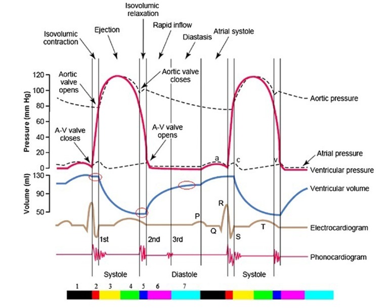
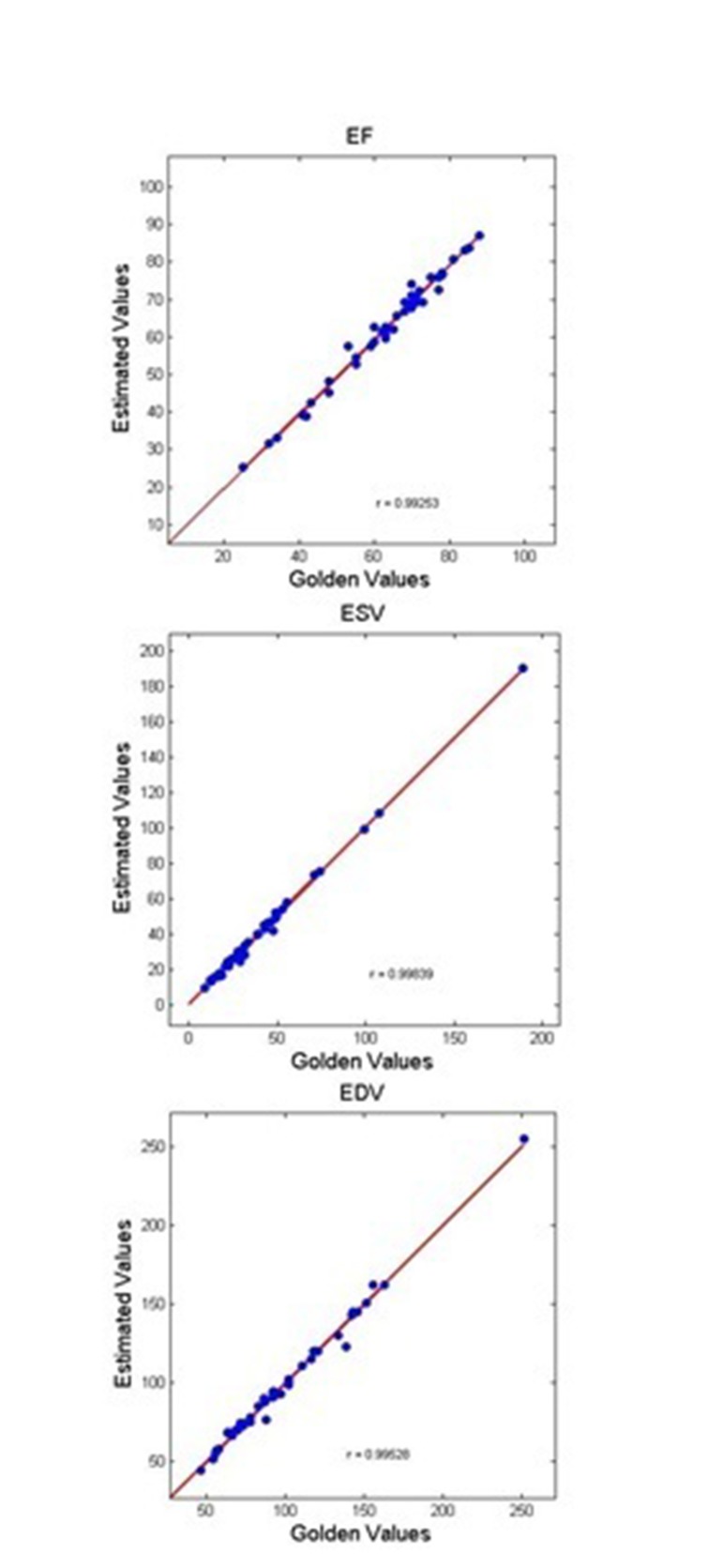
Identifying End-Diastole (ED) and End-Systole (ES) frames is highly important in the process of evaluating cardiac function and measuring global parameters accurately, such as Ejection Fraction (EF), Cardiac Output (CO) and Stroke Volume.

OBJECTIVES

The current study aimed to develop a new method based on measuring volume changes in Left Ventricle (LV) during cardiac cycle.

MATERIAL AND METHODS

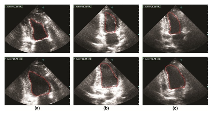
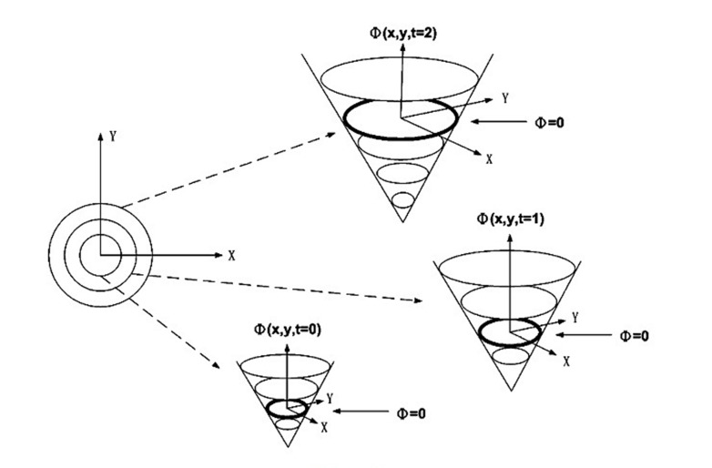
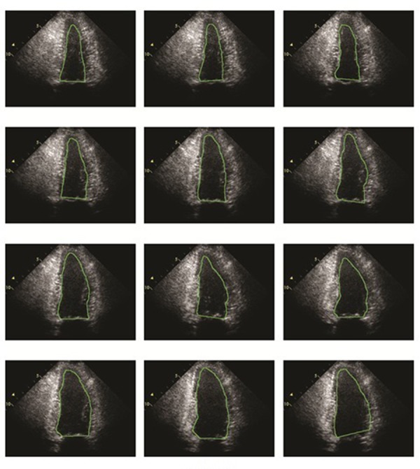
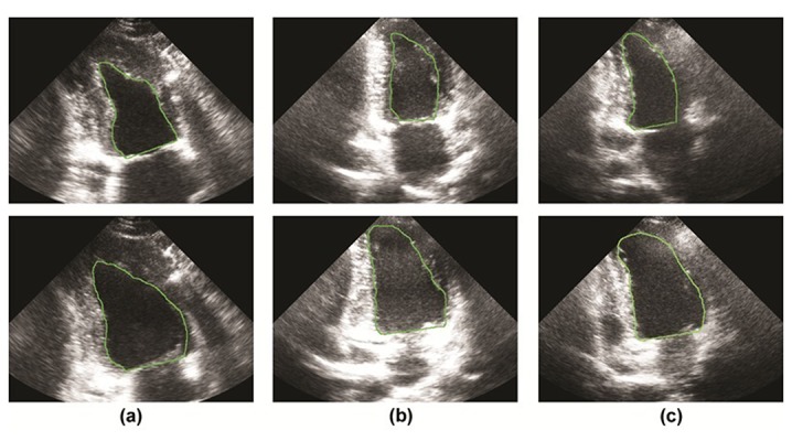
For this purpose, the Level Set method was used both in detecting endocardium border and quantifying cardiac function of all frames.

RESULTS

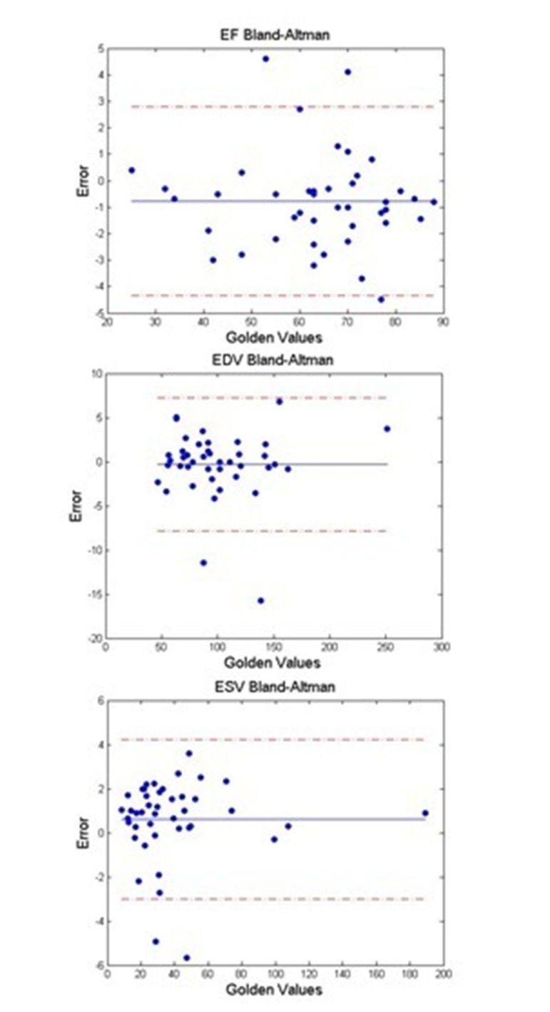
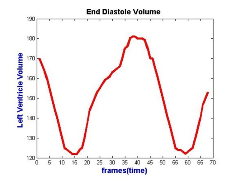
Demonstrating LV volumes displays ED and ES frames and the volumes used in calculating the required parameters.

CONCLUSIONS

Since ES and ED frames exist in iso-volumic phases of the cardiac cycle with minimum and maximum values of LV volume signals, such peaks can be utilized in finding related frames.

摘要

背景

在准确评估心脏功能和测量诸如射血分数(EF)、心输出量(CO)和每搏输出量等整体参数的过程中,识别舒张末期(ED)和收缩末期(ES)帧非常重要。

目的

本研究旨在开发一种基于测量心动周期中左心室(LV)容积变化的新方法。

材料与方法

为此,水平集方法被用于检测心内膜边界和量化所有帧的心脏功能。

结果

展示左心室容积可显示ED和ES帧以及用于计算所需参数的容积。

结论

由于ES和ED帧存在于心动周期的等容相,此时左心室容积信号具有最小值和最大值,这些峰值可用于找到相关帧。

https://cdn.ncbi.nlm.nih.gov/pmc/blobs/5bf3/4253755/060889198d7b/cardiovascmed-02-39-i005.jpg
https://cdn.ncbi.nlm.nih.gov/pmc/blobs/5bf3/4253755/3caeca3497fe/cardiovascmed-02-39-i001.jpg
https://cdn.ncbi.nlm.nih.gov/pmc/blobs/5bf3/4253755/a706e9557d21/cardiovascmed-02-39-g001.jpg
https://cdn.ncbi.nlm.nih.gov/pmc/blobs/5bf3/4253755/43a969471142/cardiovascmed-02-39-i002.jpg
https://cdn.ncbi.nlm.nih.gov/pmc/blobs/5bf3/4253755/f83e7619e5dc/cardiovascmed-02-39-i003.jpg
https://cdn.ncbi.nlm.nih.gov/pmc/blobs/5bf3/4253755/e86bce0bc129/cardiovascmed-02-39-g002.jpg
https://cdn.ncbi.nlm.nih.gov/pmc/blobs/5bf3/4253755/53d8e598d8ab/cardiovascmed-02-39-g003.jpg
https://cdn.ncbi.nlm.nih.gov/pmc/blobs/5bf3/4253755/2aa27a0a9f9f/cardiovascmed-02-39-i004.jpg
https://cdn.ncbi.nlm.nih.gov/pmc/blobs/5bf3/4253755/060889198d7b/cardiovascmed-02-39-i005.jpg

相似文献

1
Measuring Left Ventricular Volumes in Two-Dimensional Echocardiography Image Sequence Using Level-set Method for Automatic Detection of End-Diastole and End-systole Frames.使用水平集方法在二维超声心动图图像序列中测量左心室容积,以自动检测舒张末期和收缩末期帧。
Res Cardiovasc Med. 2013 Feb;2(1):39-45. doi: 10.5812/cardiovascmed.6397. Epub 2013 Feb 24.
2
Automatic detection of end-diastole and end-systole from echocardiography images using manifold learning.利用流形学习从超声心动图图像中自动检测舒张末期和收缩末期。
Physiol Meas. 2010 Sep;31(9):1091-103. doi: 10.1088/0967-3334/31/9/002. Epub 2010 Jul 23.
3
Realization of fully automated quantification of left ventricular volumes and systolic function using transthoracic 3D echocardiography.使用经胸三维超声心动图实现左心室容积和收缩功能的全自动定量分析。
Cardiovasc Ultrasound. 2018 Jan 23;16(1):2. doi: 10.1186/s12947-017-0121-8.
4
Accuracy and reproducibility of quantitation of left ventricular function by real-time three-dimensional echocardiography versus cardiac magnetic resonance.实时三维超声心动图与心脏磁共振定量左心室功能的准确性和可重复性
Am J Cardiol. 2008 Sep 15;102(6):778-83. doi: 10.1016/j.amjcard.2008.04.062. Epub 2008 Jul 9.
5
Intracardiac ultrasound measurement of volumes and ejection fraction in normal, infarcted, and aneurysmal left ventricles using a 10-MHz ultrasound catheter.使用10兆赫超声导管对正常、梗死和室壁瘤左心室进行心腔内超声测量容积和射血分数。
Circulation. 1994 Sep;90(3):1481-91. doi: 10.1161/01.cir.90.3.1481.
6
Feedback-assisted three-dimensional reconstruction of the left ventricle with MRI.磁共振成像反馈辅助的左心室三维重建
J Magn Reson Imaging. 2003 May;17(5):528-37. doi: 10.1002/jmri.10290.
7
Exploratory assessment of left ventricular strain-volume loops in severe aortic valve diseases.重度主动脉瓣疾病中左心室应变-容积环的探索性评估
J Physiol. 2017 Jun 15;595(12):3961-3971. doi: 10.1113/JP273526. Epub 2017 Mar 9.
8
Echocardiography without electrocardiogram using nonlinear dimensionality reduction methods.使用非线性降维方法的无心电图心动超声检查
J Med Ultrason (2001). 2015 Apr;42(2):137-49. doi: 10.1007/s10396-014-0588-y. Epub 2014 Nov 9.
9
[Left ventricular volume and ejection fraction by the axius auto ejection fraction method: comparison with manual trace method and visual assessment of ejection fraction].[采用Axius自动射血分数法测量左心室容积和射血分数:与手动描记法及射血分数视觉评估法的比较]
J Cardiol. 2007 Mar;49(3):125-34.
10
The impact of frame numbers on cardiac ECG-gated SPECT images with interpolated extra frames using echocardiography.使用超声心动图插入额外帧时帧数对心脏心电图门控单光子发射计算机断层扫描(SPECT)图像的影响。
Med J Islam Repub Iran. 2020 Jun 3;34:57. doi: 10.34171/mjiri.34.57. eCollection 2020.

引用本文的文献

1
CARDIAN: a novel computational approach for real-time end-diastolic frame detection in intravascular ultrasound using bidirectional attention networks.CARDIAN:一种使用双向注意力网络在血管内超声中进行实时舒张末期帧检测的新型计算方法。
Front Cardiovasc Med. 2023 Oct 6;10:1250800. doi: 10.3389/fcvm.2023.1250800. eCollection 2023.
2
Cardiac phase detection in echocardiography using convolutional neural networks.超声心动图中使用卷积神经网络进行心脏相位检测。
Sci Rep. 2023 Jun 1;13(1):8908. doi: 10.1038/s41598-023-36047-x.
3
Performance evaluation of computer-aided automated master frame selection techniques for fetal echocardiography.

本文引用的文献

1
Automatic detection of end-diastole and end-systole from echocardiography images using manifold learning.利用流形学习从超声心动图图像中自动检测舒张末期和收缩末期。
Physiol Meas. 2010 Sep;31(9):1091-103. doi: 10.1088/0967-3334/31/9/002. Epub 2010 Jul 23.
2
Incorporating temporal information into level set functional for robust ventricular boundary detection from echocardiographic image sequence.将时间信息纳入水平集函数,以从超声心动图图像序列中稳健地检测心室边界。
IEEE Trans Biomed Eng. 2008 Nov;55(11):2548-56. doi: 10.1109/TBME.2008.919135.
3
Automatic detection of end systole within a sequence of left ventricular echocardiographic images using autocorrelation and mitral valve motion detection.
计算机辅助自动主帧选择技术在胎儿超声心动图中的性能评估。
Med Biol Eng Comput. 2023 Jul;61(7):1723-1744. doi: 10.1007/s11517-023-02814-1. Epub 2023 Mar 8.
4
Automatic morphological classification of mitral valve diseases in echocardiographic images based on explainable deep learning methods.基于可解释深度学习方法的超声心动图图像中心脏二尖瓣疾病的自动形态分类。
Int J Comput Assist Radiol Surg. 2022 Feb;17(2):413-425. doi: 10.1007/s11548-021-02542-7. Epub 2021 Dec 12.
利用自相关和二尖瓣运动检测自动检测左心室超声心动图序列中的收缩末期。
Annu Int Conf IEEE Eng Med Biol Soc. 2007;2007:4504-7. doi: 10.1109/IEMBS.2007.4353340.
4
Nonlinear isochrones in murine left ventricular pressure-volume loops: how well does the time-varying elastance concept hold?小鼠左心室压力-容积环中的非线性等时线:时变弹性概念的适用程度如何?
Am J Physiol Heart Circ Physiol. 2006 Apr;290(4):H1474-83. doi: 10.1152/ajpheart.00663.2005. Epub 2005 Nov 11.
5
Assessment of left ventricular diastolic suction in dogs using wave-intensity analysis.使用波强度分析评估犬左心室舒张期抽吸功能
Am J Physiol Heart Circ Physiol. 2005 Apr;288(4):H1641-51. doi: 10.1152/ajpheart.00181.2004. Epub 2004 Nov 24.
6
Importance of mitral valve second-order chordae for left ventricular geometry, wall thickening mechanics, and global systolic function.二尖瓣二级腱索对左心室几何形态、室壁增厚力学及整体收缩功能的重要性。
Circulation. 2004 Sep 14;110(11 Suppl 1):II115-22. doi: 10.1161/01.CIR.0000138580.57971.b4.
7
Transmural mechanics at left ventricular epicardial pacing site.左心室心外膜起搏部位的透壁力学
Am J Physiol Heart Circ Physiol. 2004 Jun;286(6):H2401-7. doi: 10.1152/ajpheart.01013.2003. Epub 2004 Jan 29.
8
Effects of pulmonary insufficiency on biventricular function in the developing heart of growing swine.肺功能不全对生长猪发育中心脏双心室功能的影响。
Circulation. 2003 Oct 21;108(16):2007-13. doi: 10.1161/01.CIR.0000092887.84425.09. Epub 2003 Oct 13.
9
ACC/AHA/NASPE 2002 Guideline Update for Implantation of Cardiac Pacemakers and Antiarrhythmia Devices--summary article: a report of the American College of Cardiology/American Heart Association Task Force on Practice Guidelines (ACC/AHA/NASPE Committee to Update the 1998 Pacemaker Guidelines).ACC/AHA/NASPE 2002年心脏起搏器植入与抗心律失常器械指南更新——概述文章:美国心脏病学会/美国心脏协会实践指南工作组(ACC/AHA/NASPE 1998年起搏器指南更新委员会)报告
J Am Coll Cardiol. 2002 Nov 6;40(9):1703-19. doi: 10.1016/s0735-1097(02)02528-7.
10
A shape-space-based approach to tracking myocardial borders and quantifying regional left-ventricular function applied in echocardiography.一种基于形状空间的方法,用于在超声心动图中跟踪心肌边界并量化局部左心室功能。
IEEE Trans Med Imaging. 2002 Mar;21(3):226-38. doi: 10.1109/42.996341.