Guimarães Larissa Alves, Jimenez Paula Christine, Sousa Thiciana da Silva, Freitas Hozana Patrícia S, Rocha Danilo Damasceno, Wilke Diego Veras, Martín Jesús, Reyes Fernando, Deusdênia Loiola Pessoa Otília, Costa-Lotufo Letícia Veras
Marine Sciences Institute, Federal University of Ceara, Fortaleza, Ceara 60165-081, Brazil.
Department of Organic and Inorganic Chemistry, Federal University of Ceara, Fortaleza, CE 60021-970, Brazil.
Mar Drugs. 2014 Dec 4;12(12):5839-55. doi: 10.3390/md12125839.
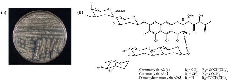
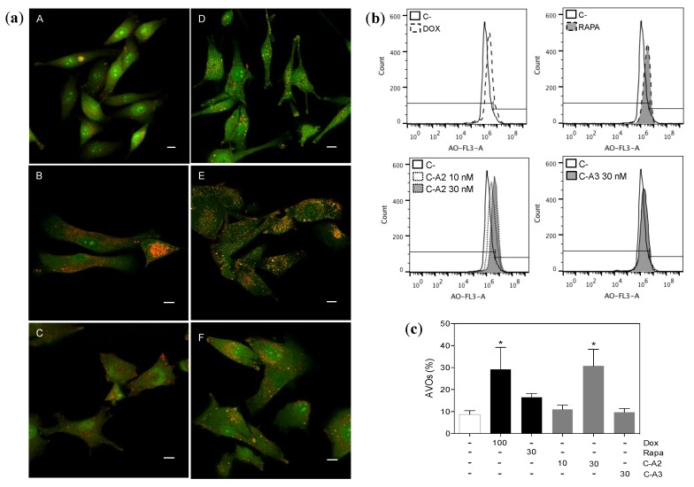
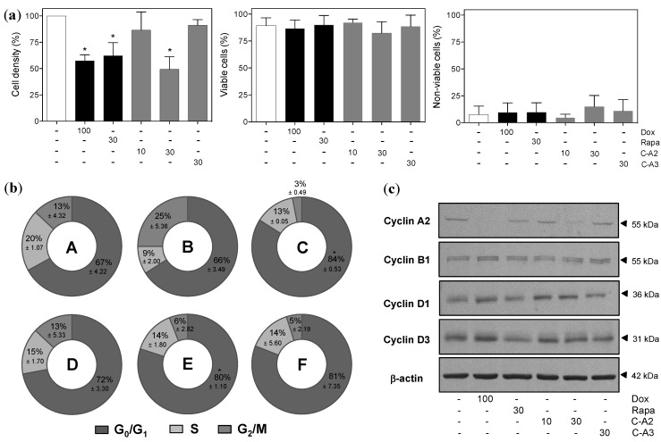
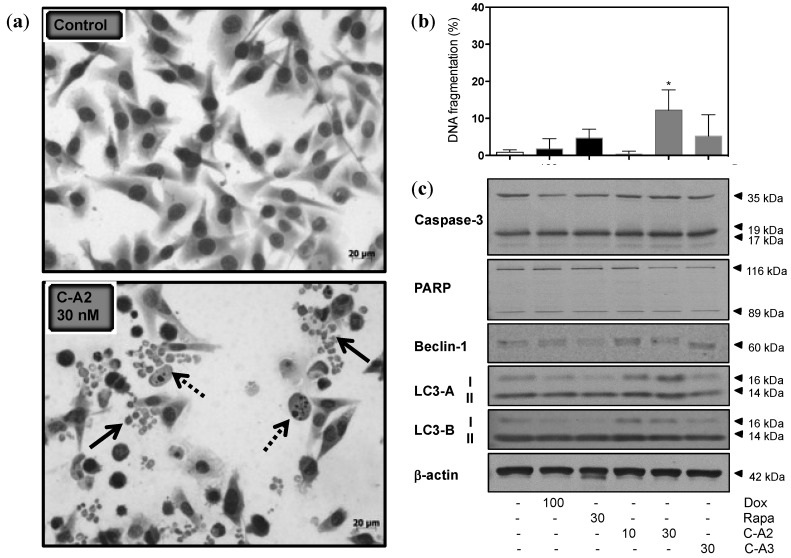
The present study highlights the biological effects of chromomycin A2 toward metastatic melanoma cells in culture. Besides chromomycin A2, chromomycin A3 and demethylchromomycin A2 were also identified from the extract derived from Streptomyces sp., recovered from Paracuru Beach, located in the northeast region of Brazil. The cytotoxic activity of chromomycin A2 was evaluated across a panel of human tumor cell lines, which found IC50 values in the nM-range for exposures of 48 and 72 h. MALME-3M, a metastatic melanoma cell line, showed the highest sensitivity to chromomycin A2 after 48h incubation, and was chosen as a model to investigate this potent cytotoxic effect. Treatment with chromomycin A2 at 30 nM reduced cell proliferation, but had no significant effect upon cell viability. Additionally, chromomycin A2 induced accumulation of cells in G0/G1 phase of the cell cycle, with consequent reduction of S and G2/M and unbalanced expression of cyclins. Chromomycin A2 treated cells depicted several cellular fragments resembling autophagosomes and increased expression of proteins LC3-A and LC3-B. Moreover, exposure to chromomycin A2 also induced the appearance of acidic vacuolar organelles in treated cells. These features combined are suggestive of the induction of autophagy promoted by chromomycin A2, a feature not previously described for chromomycins.
本研究着重探讨了放线菌素A2对培养的转移性黑色素瘤细胞的生物学效应。除了放线菌素A2外,还从巴西东北部帕拉库鲁海滩分离出的链霉菌提取物中鉴定出了放线菌素A3和去甲基放线菌素A2。在一组人类肿瘤细胞系中评估了放线菌素A2的细胞毒性活性,结果发现在48小时和72小时暴露时,其IC50值在纳摩尔范围内。转移性黑色素瘤细胞系MALME-3M在孵育48小时后对放线菌素A2表现出最高的敏感性,并被选作模型来研究这种强大的细胞毒性作用。用30 nM的放线菌素A2处理可降低细胞增殖,但对细胞活力没有显著影响。此外,放线菌素A2诱导细胞在细胞周期的G0/G1期积累,从而导致S期和G2/M期减少以及细胞周期蛋白表达失衡。经放线菌素A2处理的细胞呈现出几个类似于自噬体的细胞碎片,并增加了LC3-A和LC3-B蛋白的表达。此外,暴露于放线菌素A2还诱导处理后的细胞中出现酸性液泡细胞器。这些特征综合起来表明放线菌素A2可诱导自噬,这是以前未在放线菌素中描述过的特征。