• 文献检索
  • 文档翻译
  • 深度研究
  • 学术资讯
  • Suppr Zotero 插件Zotero 插件
  • 邀请有礼
  • 套餐&价格
  • 历史记录
应用&插件
Suppr Zotero 插件Zotero 插件浏览器插件Mac 客户端Windows 客户端微信小程序
定价
高级版会员购买积分包购买API积分包
服务
文献检索文档翻译深度研究API 文档MCP 服务
关于我们
关于 Suppr公司介绍联系我们用户协议隐私条款
关注我们

Suppr 超能文献

核心技术专利:CN118964589B侵权必究
粤ICP备2023148730 号-1Suppr @ 2026

文献检索

告别复杂PubMed语法,用中文像聊天一样搜索,搜遍4000万医学文献。AI智能推荐,让科研检索更轻松。

立即免费搜索

文件翻译

保留排版,准确专业,支持PDF/Word/PPT等文件格式,支持 12+语言互译。

免费翻译文档

深度研究

AI帮你快速写综述,25分钟生成高质量综述,智能提取关键信息,辅助科研写作。

立即免费体验

在神经外科重症监护病房患者中使用超声评估脑中线移位

Assessment of brain midline shift using sonography in neurosurgical ICU patients.

作者信息

Motuel Julie, Biette Isaure, Srairi Mohamed, Mrozek Ségolène, Kurrek Matt M, Chaynes Patrick, Cognard Christophe, Fourcade Olivier, Geeraerts Thomas

机构信息

Anesthesiology and Critical Care Department, Equipe d'accueil "Modélisation de l'agression tissulaire et nociceptive", University Hospital of Toulouse, University Toulouse 3 Paul Sabatier, Toulouse, France.

Neuroradiology Department, University Hospital of Toulouse, University Toulouse 3 Paul Sabatier, Toulouse, France.

出版信息

Crit Care. 2014 Dec 9;18(6):676. doi: 10.1186/s13054-014-0676-9.

DOI:10.1186/s13054-014-0676-9
PMID:25488604
原文链接:https://pmc.ncbi.nlm.nih.gov/articles/PMC4305234/
Abstract

INTRODUCTION

Brain midline shift (MLS) is a life-threatening condition that requires urgent diagnosis and treatment. We aimed to validate bedside assessment of MLS with Transcranial Sonography (TCS) in neurosurgical ICU patients by comparing it to CT.

METHODS

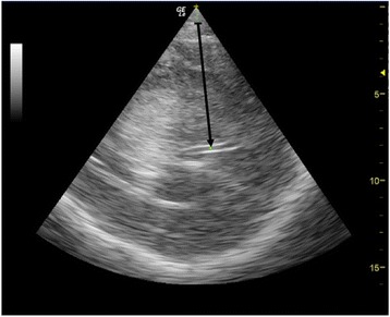
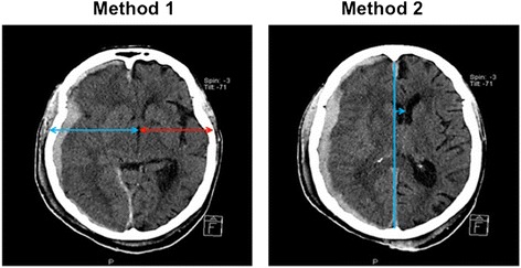
In this prospective single centre study, patients who underwent a head CT were included and a concomitant TCS performed. TCS MLS was determined by measuring the difference between the distance from skull to the third ventricle on both sides, using a 2 to 4 MHz probe through the temporal window. CT MLS was measured as the difference between the ideal midline and the septum pellucidum. A significant MLS was defined on head CT as > 0.5 cm.

RESULTS

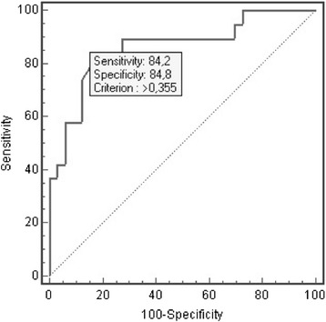
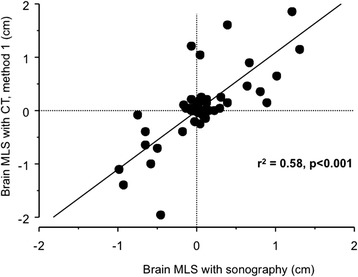
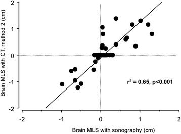
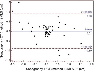
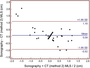
A total of 52 neurosurgical ICU patients were included. The MLS (mean ± SD) was 0.32 ± 0.36 cm using TCS and 0.47 ± 0.67 cm using CT. The Pearson's correlation coefficient (r(2)) between TCS and CT scan was 0.65 (P < 0.001). The bias was 0.09 cm and the limits of agreements were 1.10 and -0.92 cm. The area under the ROC curve for detecting a significant MLS with TCS was 0.86 (95% CI = 0.74 to 0.94), and, using 0.35 cm as a cut-off, the sensitivity was 84.2%, the specificity 84.8% and the positive likelihood ratio was 5.56.

CONCLUSIONS

This study suggests that TCS could detect MLS with reasonable accuracy in neurosurgical ICU patients and that it could serve as a bedside tool to facilitate early diagnosis and treatment for patients with a significant intracranial mass effect.

摘要

引言

脑中线移位(MLS)是一种危及生命的状况,需要紧急诊断和治疗。我们旨在通过将经颅超声检查(TCS)与CT进行比较,验证神经外科重症监护病房(ICU)患者床边MLS评估的准确性。

方法

在这项前瞻性单中心研究中,纳入接受头部CT检查的患者,并同时进行TCS检查。使用2至4MHz探头通过颞窗测量两侧颅骨至第三脑室距离的差值来确定TCS MLS。CT MLS测量为理想中线与透明隔之间的差值。头部CT上显著MLS定义为>0.5cm。

结果

共纳入52例神经外科ICU患者。TCS测量的MLS(均值±标准差)为0.32±0.36cm,CT测量的为0.47±0.67cm。TCS与CT扫描之间的Pearson相关系数(r(2))为0.65(P<0.001)。偏差为0.09cm,一致性界限为1.10和-0.92cm。TCS检测显著MLS的ROC曲线下面积为0.86(95%CI=0.74至0.94),以0.35cm为临界值时,敏感性为84.2%,特异性为84.8%,阳性似然比为5.56。

结论

本研究表明,TCS能够在神经外科ICU患者中以合理的准确性检测MLS,并且可作为一种床边工具,便于对有显著颅内占位效应的患者进行早期诊断和治疗。

https://cdn.ncbi.nlm.nih.gov/pmc/blobs/3586/4305234/70cf674ca103/13054_2014_676_Fig7_HTML.jpg
https://cdn.ncbi.nlm.nih.gov/pmc/blobs/3586/4305234/ed545bedb529/13054_2014_676_Fig1_HTML.jpg
https://cdn.ncbi.nlm.nih.gov/pmc/blobs/3586/4305234/6b6bf60f928c/13054_2014_676_Fig2_HTML.jpg
https://cdn.ncbi.nlm.nih.gov/pmc/blobs/3586/4305234/22e90ed4fa89/13054_2014_676_Fig3_HTML.jpg
https://cdn.ncbi.nlm.nih.gov/pmc/blobs/3586/4305234/1b401a483a26/13054_2014_676_Fig4_HTML.jpg
https://cdn.ncbi.nlm.nih.gov/pmc/blobs/3586/4305234/41e364567618/13054_2014_676_Fig5_HTML.jpg
https://cdn.ncbi.nlm.nih.gov/pmc/blobs/3586/4305234/1fdcf933b370/13054_2014_676_Fig6_HTML.jpg
https://cdn.ncbi.nlm.nih.gov/pmc/blobs/3586/4305234/70cf674ca103/13054_2014_676_Fig7_HTML.jpg

相似文献

1
Assessment of brain midline shift using sonography in neurosurgical ICU patients.在神经外科重症监护病房患者中使用超声评估脑中线移位
Crit Care. 2014 Dec 9;18(6):676. doi: 10.1186/s13054-014-0676-9.
2
Accuracy of bedside bidimensional transcranial ultrasound versus tomodensitometric measurement of the third ventricle.床边二维经颅超声与断层扫描测量第三脑室准确性的比较。
J Neuroimaging. 2022 Jul;32(4):629-637. doi: 10.1111/jon.12970. Epub 2022 Jan 26.
3
Transcranial color-coded duplex sonography for evaluation of midline-shift after chronic-subdural hematoma evacuation (TEMASE): A prospective study.经颅彩色编码双功超声评估慢性硬膜下血肿清除术后中线移位(TEMASE):一项前瞻性研究
Clin Neurol Neurosurg. 2017 Nov;162:101-107. doi: 10.1016/j.clineuro.2017.09.015. Epub 2017 Oct 3.
4
Comparison between Transcranial Sonography and Computerized Tomography Scans to Assess the Midline Shift in Patients with Traumatic Brain Injury.经颅超声检查与计算机断层扫描在评估创伤性脑损伤患者中线移位中的比较
Indian J Crit Care Med. 2023 Jan;27(1):64-66. doi: 10.5005/jp-journals-10071-24376.
5
Application of Transcranial Sonography for the Assessment of Brain Midline Shift in Patients Presenting With Suspected Intracranial Pathology to the Emergency Department of a Tertiary Care Hospital in Central Gujarat, India.经颅超声检查在印度古吉拉特邦中部一家三级护理医院急诊科疑似颅内病变患者脑中线移位评估中的应用
Cureus. 2024 Jan 19;16(1):e52561. doi: 10.7759/cureus.52561. eCollection 2024 Jan.
6
Assessment of Midline Shift in Postdecompressive Craniectomy Patients in Neurocritical Care: Comparison between Transcranial Ultrasonography and Computerized Tomography.神经重症监护中去骨瓣减压术后中线移位的评估:经颅超声与计算机断层扫描的比较。
Neurol India. 2023 Nov-Dec;71(6):1167-1171. doi: 10.4103/0028-3886.391386.
7
Correlation between Transcranial Ultrasound and CT Head to Detect Clinically Significant Conditions in Post-craniectomy Patients Performed by Emergency Physician: A Pilot Study.急诊医生对颅骨切除术后患者经颅超声与头颅CT检测临床重要情况的相关性:一项初步研究
Indian J Crit Care Med. 2024 Mar;28(3):299-306. doi: 10.5005/jp-journals-10071-24662.
8
Monitoring midline shift by transcranial color-coded sonography in traumatic brain injury. A comparison with cranial computerized tomography.
Intensive Care Med. 2004 Aug;30(8):1672-5. doi: 10.1007/s00134-004-2348-8. Epub 2004 Jun 9.
9
Third ventricle midline shift due to spontaneous supratentorial intracerebral hemorrhage evaluated by transcranial color-coded sonography.经颅彩色编码超声评估自发性幕上脑出血所致第三脑室中线移位
J Ultrasound Med. 2006 Feb;25(2):203-9. doi: 10.7863/jum.2006.25.2.203.
10
Transcranial sonographic monitoring of hemorrhagic transformation in patients with acute middle cerebral artery infarction.急性大脑中动脉梗死患者出血性转化的经颅超声监测
J Neuroimaging. 2005 Oct;15(4):326-30. doi: 10.1177/1051228405280174.

引用本文的文献

1
Management of intracranial hypertension with and without invasive intracranial pressure monitoring.有创和无创颅内压监测下的颅内高压管理
World J Crit Care Med. 2025 Sep 9;14(3):105645. doi: 10.5492/wjccm.v14.i3.105645.
2
A New Predictor Score for Postoperative Seizures in Brain Tumor Patients Without a Seizure History (BRAINNN Score).一种用于无癫痫病史的脑肿瘤患者术后癫痫发作的新预测评分(BRAINNN评分)。
Cureus. 2025 May 19;17(5):e84424. doi: 10.7759/cureus.84424. eCollection 2025 May.
3
Hocus pocus: advanced point-of-care ultrasound from the trauma bay to the surgical ICU.

本文引用的文献

1
Sonographic monitoring of mass effect in stroke patients treated with hypothermia. Correlation with intracranial pressure and matrix metalloproteinase 2 and 9 expression.超声监测低温治疗的中风患者的占位效应。与颅内压及基质金属蛋白酶2和9表达的相关性。
J Neurol Sci. 2009 Jan 15;276(1-2):75-8. doi: 10.1016/j.jns.2008.08.038. Epub 2008 Oct 2.
2
Predicting outcome after traumatic brain injury: practical prognostic models based on large cohort of international patients.预测创伤性脑损伤后的结局:基于大量国际患者队列的实用预后模型。
BMJ. 2008 Feb 23;336(7641):425-9. doi: 10.1136/bmj.39461.643438.25. Epub 2008 Feb 12.
3
变戏法:从创伤病房到外科重症监护病房的高级床旁超声检查
Trauma Surg Acute Care Open. 2025 Apr 14;10(Suppl 1):e001779. doi: 10.1136/tsaco-2025-001779. eCollection 2025.
4
Identifying research activity on brain ultrasonography in craniocerebral diseases by bibliometric and visualized analysis of a 20-year journey of global publications.通过对20年全球出版物历程的文献计量学和可视化分析,确定颅脑疾病中脑超声检查的研究活动。
Medicine (Baltimore). 2025 Mar 21;104(12):e41927. doi: 10.1097/MD.0000000000041927.
5
Transcranial sonography: practical use in the intensive care unit.经颅超声检查:在重症监护病房的实际应用
Anaesthesiol Intensive Ther. 2024;56(5):267-276. doi: 10.5114/ait.2024.146640.
6
Non-Invasive Monitoring of Cerebral Edema Using Ultrasonic Echo Signal Features and Machine Learning.利用超声回波信号特征和机器学习对脑水肿进行无创监测
Brain Sci. 2024 Nov 23;14(12):1175. doi: 10.3390/brainsci14121175.
7
Application of Transcranial Sonography for the Assessment of Brain Midline Shift in Patients Presenting With Suspected Intracranial Pathology to the Emergency Department of a Tertiary Care Hospital in Central Gujarat, India.经颅超声检查在印度古吉拉特邦中部一家三级护理医院急诊科疑似颅内病变患者脑中线移位评估中的应用
Cureus. 2024 Jan 19;16(1):e52561. doi: 10.7759/cureus.52561. eCollection 2024 Jan.
8
Head to toe ultrasound: a narrative review of experts' recommendations of methodological approaches.从头到脚超声检查:专家关于方法学方法建议的叙述性综述
J Anesth Analg Crit Care. 2022 Oct 21;2(1):44. doi: 10.1186/s44158-022-00072-5.
9
POCUS, how can we include the brain? An overview.床旁超声检查,我们如何将脑部纳入其中?概述。
J Anesth Analg Crit Care. 2022 Dec 27;2(1):55. doi: 10.1186/s44158-022-00082-3.
10
Comparison between Transcranial Sonography and Computerized Tomography Scans to Assess the Midline Shift in Patients with Traumatic Brain Injury.经颅超声检查与计算机断层扫描在评估创伤性脑损伤患者中线移位中的比较
Indian J Crit Care Med. 2023 Jan;27(1):64-66. doi: 10.5005/jp-journals-10071-24376.
Surgical management of acute subdural hematomas.
急性硬膜下血肿的外科治疗
Neurosurgery. 2006 Mar;58(3 Suppl):S16-24; discussion Si-iv.
4
Surgical management of acute epidural hematomas.急性硬膜外血肿的手术治疗
Neurosurgery. 2006 Mar;58(3 Suppl):S7-15; discussion Si-iv.
5
Surgical management of traumatic parenchymal lesions.创伤性实质病变的外科治疗
Neurosurgery. 2006 Mar;58(3 Suppl):S25-46; discussion Si-iv. doi: 10.1227/01.NEU.0000210365.36914.E3.
6
Third ventricle midline shift due to spontaneous supratentorial intracerebral hemorrhage evaluated by transcranial color-coded sonography.经颅彩色编码超声评估自发性幕上脑出血所致第三脑室中线移位
J Ultrasound Med. 2006 Feb;25(2):203-9. doi: 10.7863/jum.2006.25.2.203.
7
Prediction of outcome in traumatic brain injury with computed tomographic characteristics: a comparison between the computed tomographic classification and combinations of computed tomographic predictors.利用计算机断层扫描特征预测创伤性脑损伤的预后:计算机断层扫描分类与计算机断层扫描预测指标组合之间的比较
Neurosurgery. 2005 Dec;57(6):1173-82; discussion 1173-82. doi: 10.1227/01.neu.0000186013.63046.6b.
8
Monitoring midline shift by transcranial color-coded sonography in traumatic brain injury. A comparison with cranial computerized tomography.
Intensive Care Med. 2004 Aug;30(8):1672-5. doi: 10.1007/s00134-004-2348-8. Epub 2004 Jun 9.
9
Routinely repeated computed tomography after blunt head trauma: does it benefit patients?钝性头部创伤后常规重复计算机断层扫描:对患者有益吗?
J Trauma. 2004 Mar;56(3):475-80; discussion 480-1. doi: 10.1097/01.ta.0000114304.56006.d4.
10
Sonographic monitoring of midline shift in space-occupying stroke: an early outcome predictor.
Stroke. 2001 Feb;32(2):442-7. doi: 10.1161/01.str.32.2.442.