Zhang Wei, Thamattoor Ajoy K, LeRoy Christopher, Buckmaster Paul S
Department of Comparative Medicine, Stanford University, Stanford, California.
Hippocampus. 2015 May;25(5):594-604. doi: 10.1002/hipo.22396. Epub 2014 Dec 26.
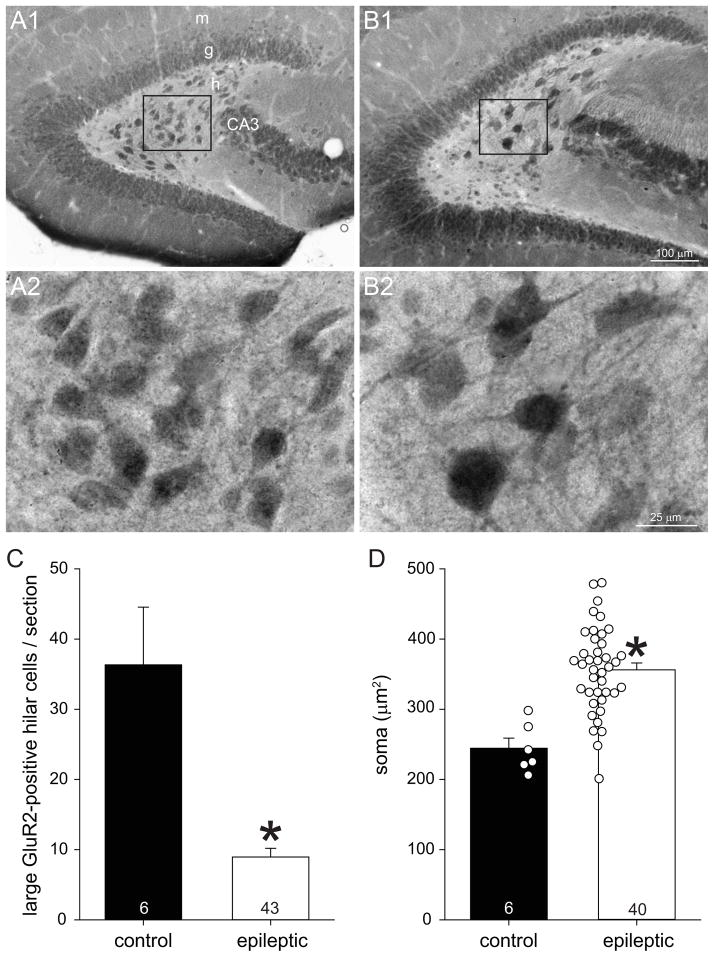
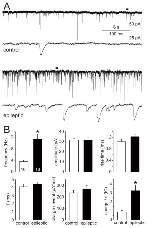
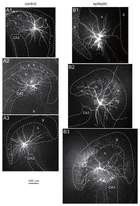
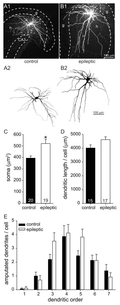
Numerous hypotheses of temporal lobe epileptogenesis have been proposed, and several involve hippocampal mossy cells. Building on previous hypotheses we sought to test the possibility that after epileptogenic injuries surviving mossy cells develop into super-connected seizure-generating hub cells. If so, they might require more cellular machinery and consequently have larger somata, elongate their dendrites to receive more synaptic input, and display higher frequencies of miniature excitatory synaptic currents (mEPSCs). To test these possibilities pilocarpine-treated mice were evaluated using GluR2-immunocytochemistry, whole-cell recording, and biocytin-labeling. Epileptic pilocarpine-treated mice displayed substantial loss of GluR2-positive hilar neurons. Somata of surviving neurons were 1.4-times larger than in controls. Biocytin-labeled mossy cells also were larger in epileptic mice, but dendritic length per cell was not significantly different. The average frequency of mEPSCs of mossy cells recorded in the presence of tetrodotoxin and bicuculline was 3.2-times higher in epileptic pilocarpine-treated mice as compared to controls. Other parameters of mEPSCs were similar in both groups. Average input resistance of mossy cells in epileptic mice was reduced to 63% of controls, which is consistent with larger somata and would tend to make surviving mossy cells less excitable. Other intrinsic physiological characteristics examined were similar in both groups. Increased excitatory synaptic input is consistent with the hypothesis that surviving mossy cells develop into aberrantly super-connected seizure-generating hub cells, and soma hypertrophy is indirectly consistent with the possibility of axon sprouting. However, no obvious evidence of hyperexcitable intrinsic physiology was found. Furthermore, similar hypertrophy and hyper-connectivity has been reported for other neuron types in the dentate gyrus, suggesting mossy cells are not unique in this regard. Thus, findings of the present study reveal epilepsy-related changes in mossy cell anatomy and synaptic input but do not strongly support the hypothesis that mossy cells develop into seizure-generating hub cells.
人们已经提出了许多关于颞叶癫痫发生机制的假说,其中一些涉及海马苔藓细胞。基于先前的假说,我们试图检验以下可能性:在致痫性损伤后,存活的苔藓细胞是否会发展成为超连接的癫痫发作起始枢纽细胞。如果是这样,它们可能需要更多的细胞机制,因此具有更大的胞体,延长其树突以接收更多的突触输入,并表现出更高频率的微小兴奋性突触电流(mEPSCs)。为了验证这些可能性,我们使用GluR2免疫细胞化学、全细胞记录和生物素标记对毛果芸香碱处理的小鼠进行了评估。癫痫性毛果芸香碱处理的小鼠显示GluR2阳性的门区神经元大量丧失。存活神经元的胞体比对照组大1.4倍。生物素标记的苔藓细胞在癫痫小鼠中也更大,但每个细胞的树突长度没有显著差异。在存在河豚毒素和荷包牡丹碱的情况下,癫痫性毛果芸香碱处理的小鼠中记录到的苔藓细胞mEPSCs平均频率比对照组高3.2倍。两组mEPSCs的其他参数相似。癫痫小鼠中苔藓细胞的平均输入电阻降至对照组的63%,这与更大的胞体一致,并且倾向于使存活的苔藓细胞兴奋性降低。所检查的其他内在生理特征在两组中相似。兴奋性突触输入增加与存活的苔藓细胞发展成为异常超连接的癫痫发作起始枢纽细胞的假说一致,胞体肥大间接与轴突发芽的可能性一致。然而,未发现明显的内在生理兴奋性过高的证据。此外,已报道齿状回中其他神经元类型也有类似的肥大和超连接现象,这表明苔藓细胞在这方面并非独一无二。因此,本研究结果揭示了苔藓细胞解剖结构和突触输入中与癫痫相关的变化,但并不强烈支持苔藓细胞发展成为癫痫发作起始枢纽细胞的假说。