Department of Pharmacology, University of Oxford Mansfield Road, Oxford, OX1 3QT, United Kingdom.
F. Hoffmann-La Roche Ltd. Building 74/3W.306A, Grenzacherstrasse 183, CH-4070, Basel, Switzerland.
Ann Clin Transl Neurol. 2014 Sep;1(9):659-69. doi: 10.1002/acn3.94. Epub 2014 Sep 16.
The mechanism of action of anti-B cell therapy in multiple sclerosis (MS) is not fully understood. Here, we compared the effect of anti-CD20 therapy on microglial activation in two distinct focal rat models of MS.
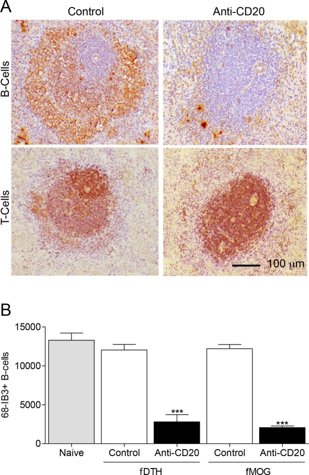
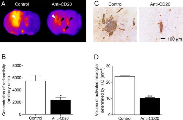
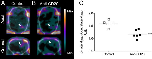
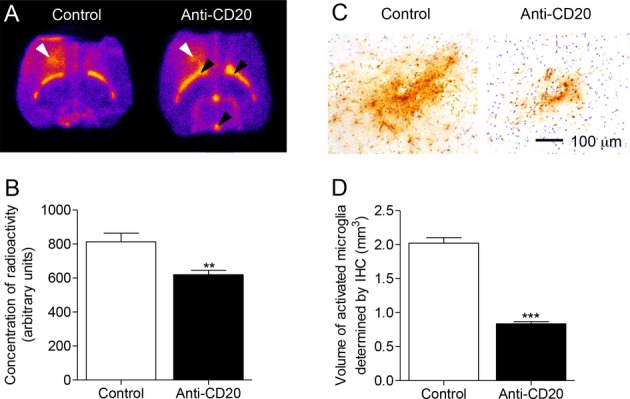
The effect of anti-CD20 therapy on lesion formation and extralesional microglial activation was evaluated in the fDTH-EAE (experimental allergic encephalomyelitis) model, which is a focal demyelinating type-IV delayed-type hypersensitivity lesion. For comparison, effects were also assessed in the focal humoral MOG model induced by intracerebral injection of cytokine in myelin oligodendrocyte glycoprotein immunized rats. Microglial activation was assessed in situ and in vivo using the TSPO SPECT ligand [(125)I]DPA-713, and by immunostaining for MHCII. The effect of treatment on demyelination and lymphocyte recruitment to the brain were evaluated.
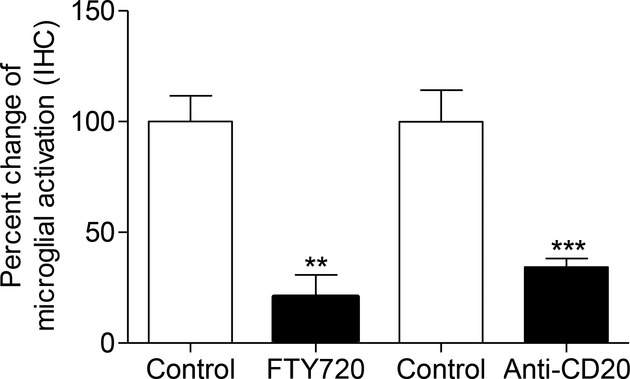
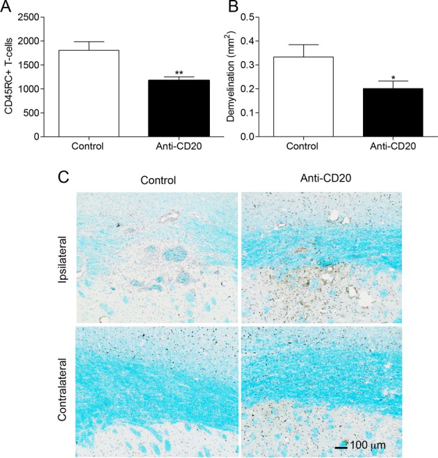
Anti-CD20 therapy reduced microglial activation, and lesion formation in the humoral model, but it was most effective in the antibody-independent fDTH-EAE. Immunohistochemistry for MHCII also demonstrated a reduced volume of microglial activation in the brains of anti-CD20-treated fDTH-EAE animals, which was accompanied by a reduction in T-cell recruitment and demyelination. The effect anti-CD20 therapy in the latter model was similarly strong as compared to the T-cell targeting MS compound FTY720.
The suppression of lesion development by anti-CD20 treatment in an antibody-independent model suggests that B-cells play an important role in lesion development, independent of auto-antibody production. Thus, CD20-positive B-cell depletion has the potential to be effective in a wider population of individuals with MS than might have been predicted from our knowledge of the underlying histopathology.
抗 B 细胞治疗在多发性硬化症(MS)中的作用机制尚未完全阐明。在这里,我们比较了抗 CD20 治疗对两种不同的 MS 局灶性大鼠模型中小胶质细胞激活的影响。
在实验性过敏性脑脊髓炎(EAE)模型(一种局灶性脱髓鞘 IV 型迟发型超敏反应病变)中,评估了抗 CD20 治疗对病变形成和病变外小胶质细胞激活的影响。为了进行比较,还评估了在细胞因子脑内注射诱导的局灶性体液性 MOG 模型中的作用。使用 TSPO SPECT 配体 [(125)I]DPA-713 以及 MHCII 免疫染色,原位和体内评估小胶质细胞激活。评估治疗对脱髓鞘和淋巴细胞向脑内募集的影响。
抗 CD20 治疗减少了体液模型中的小胶质细胞激活和病变形成,但在抗体非依赖性 fDTH-EAE 中效果最为显著。抗 CD20 处理 fDTH-EAE 动物的大脑中 MHCII 的免疫组织化学也表明小胶质细胞激活体积减少,伴随 T 细胞募集和脱髓鞘减少。与 T 细胞靶向 MS 化合物 FTY720 相比,后一种模型中抗 CD20 治疗的效果同样强烈。
抗 CD20 治疗在抗体非依赖性模型中抑制病变发展表明 B 细胞在病变发展中发挥重要作用,而与自身抗体产生无关。因此,CD20 阳性 B 细胞耗竭有可能比我们对潜在组织病理学的了解所预测的更广泛地应用于多发性硬化症患者。