Vainio S K, Dickens A M, Matilainen M, López-Picón F R, Aarnio R, Eskola O, Solin O, Anthony D C, Rinne J O, Airas L, Haaparanta-Solin M
Turku PET Centre, Preclinical PET Imaging, Preclinical Imaging Laboratory, University of Turku, Tykistökatu 6 A, 20520, Turku, Finland.
MediCity Research Laboratory, University of Turku, Turku, Finland.
EJNMMI Res. 2022 Feb 2;12(1):6. doi: 10.1186/s13550-022-00878-y.
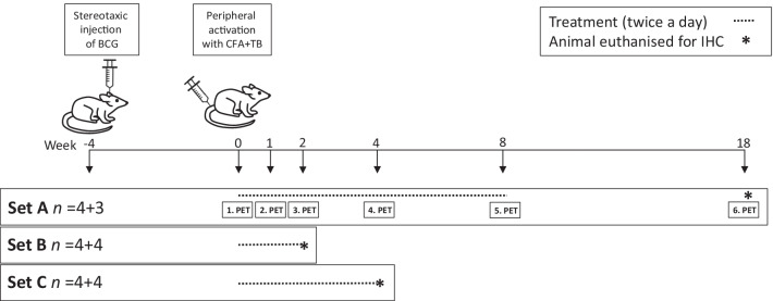
Dimethyl fumarate (DMF) is an oral immunomodulatory drug used in the treatment of autoimmune diseases. Here, we sought to study whether the effect of DMF can be detected using positron emission tomography (PET) targeting the 18-kDa translocator protein (TSPO) in the focal delayed-type hypersensitivity rat model of multiple sclerosis (fDTH-EAE). The rats were treated orally twice daily from lesion activation (day 0) with either vehicle (tap water with 0.08% Methocel, 200 µL; control group n = 4 (3 after week four)) or 15 mg/kg DMF (n = 4) in 0.08% aqueous Methocel (200 µL) for 8 weeks. The animals were imaged by PET using the TSPO tracer [F]GE-180 in weeks 0, 1, 2, 4, 8, and 18 following lesion activation, and the non-displaceable binding potential (BP) was calculated. Immunohistochemical staining for Iba1, CD4, and CD8 was performed in week 18, and in separate cohorts of animals, following 2 or 4 weeks of treatment.
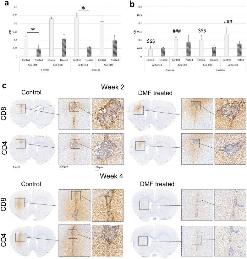
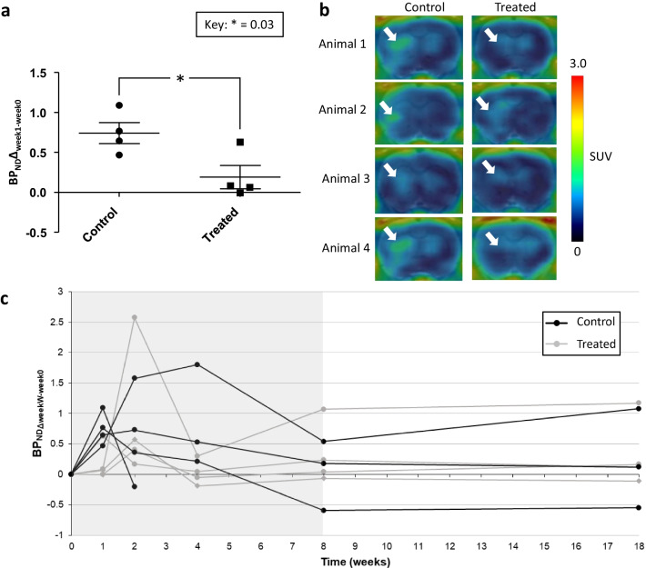
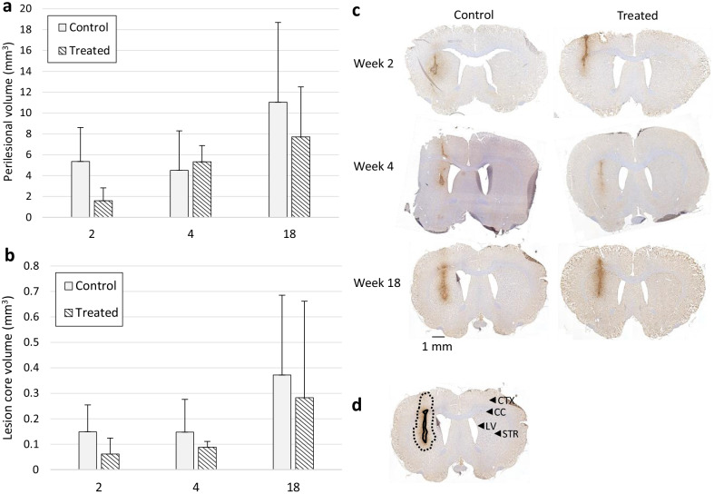
Using the fDTH-EAE model, DMF reduced the [F]GE-180 BP in the DMF-treated animals compared to control animals after 1 week of treatment (two-tailed unpaired t test, p = 0.031), but not in weeks 2, 4, 8, or 18 when imaged in vivo by PET. Immunostaining for Iba1 showed that DMF had no effect on the perilesional volume or the core lesion volume after 2 or 4 weeks of treatment, or at 18 weeks. However, the optical density (OD) measurements of CD4 staining showed reduced OD in the lesions of the treated rats.
DMF reduced the microglial activation in the fDTH-EAE model after 1 week of treatment, as detected by PET imaging of the TSPO ligand [F]GE-180. However, over an extended time course, reduced microglial activation was not observed using [F]GE-180 or by immunohistochemistry for Iba1 microglia/macrophages. Additionally, DMF did affect the infiltration of CD4 and CD8 T-lymphocytes at the fDTH-EAE lesion.
富马酸二甲酯(DMF)是一种用于治疗自身免疫性疾病的口服免疫调节药物。在此,我们试图研究在多发性硬化症局灶性迟发型超敏反应大鼠模型(fDTH-EAE)中,是否可以使用正电子发射断层扫描(PET)靶向18 kDa转位蛋白(TSPO)来检测DMF的效果。从病变激活(第0天)开始,大鼠每天口服两次,对照组给予赋形剂(含0.08%甲基纤维素的自来水,200 μL;n = 4只(四周后3只)),DMF组给予15 mg/kg DMF(n = 4只),溶于0.08%甲基纤维素水溶液(200 μL)中,持续8周。在病变激活后的第0、1、2、4、8和18周,使用TSPO示踪剂[F]GE-180对动物进行PET成像,并计算非置换性结合潜能(BP)。在第18周以及在单独的动物组中,在治疗2周或4周后,对Iba1、CD4和CD8进行免疫组织化学染色。
使用fDTH-EAE模型,与对照组动物相比,DMF治疗1周后降低了DMF治疗组动物的[F]GE-180 BP(双侧不成对t检验,p = 0.031),但在第2、4、8或18周通过PET进行体内成像时未降低。Iba1免疫染色显示,DMF在治疗2周或4周后以及18周时,对病变周围体积或核心病变体积没有影响。然而,CD4染色的光密度(OD)测量显示,治疗大鼠病变中的OD降低。
通过TSPO配体[F]GE-180的PET成像检测,DMF在治疗1周后降低了fDTH-EAE模型中的小胶质细胞活化。然而,在较长时间过程中,使用[F]GE-180或通过Iba1小胶质细胞/巨噬细胞的免疫组织化学未观察到小胶质细胞活化降低。此外,DMF确实影响了fDTH-EAE病变处CD4和CD8 T淋巴细胞的浸润。