Mao Yan, Qu Qing, Zhang Yuzi, Liu Junjun, Chen Xiaosong, Shen Kunwei
Comprehensive Breast Health Center, Ruijin Hospital, Shanghai Jiao Tong University School of Medicine, Shanghai, China.
Department of Oncology, Ruijin Hospital, Shanghai Jiao Tong University School of Medicine, Shanghai, China.
PLoS One. 2014 Dec 12;9(12):e115103. doi: 10.1371/journal.pone.0115103. eCollection 2014.
We carried out a systematic review and meta-analysis to evaluate the predictive roles of tumor infiltrating lymphocytes (TILs) in response to neoadjuvant chemotherapy (NAC) in breast cancer.
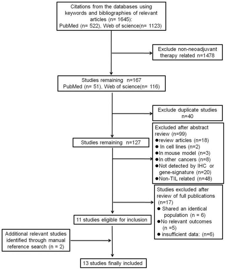
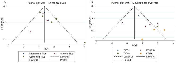
A PubMed and Web of Science literature search was designed. Random or fixed effect models were adopted to estimate the summary odds ratio (OR). Heterogeneity and sensitivity analyses were performed to explore heterogeneity among studies and to assess the effects of study quality. Publication bias was evaluated using a funnel plot, Egger's test and Begg's test. We included studies where the predictive significance of TILs, and/or TILs subset on the pathologic complete response (pCR) were determined in NAC of breast cancer.
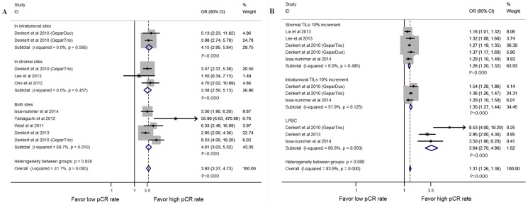
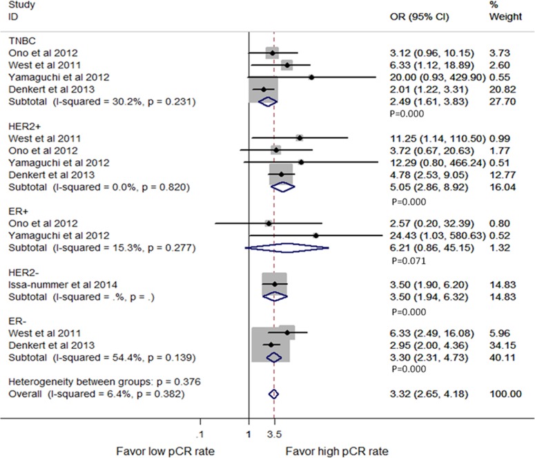
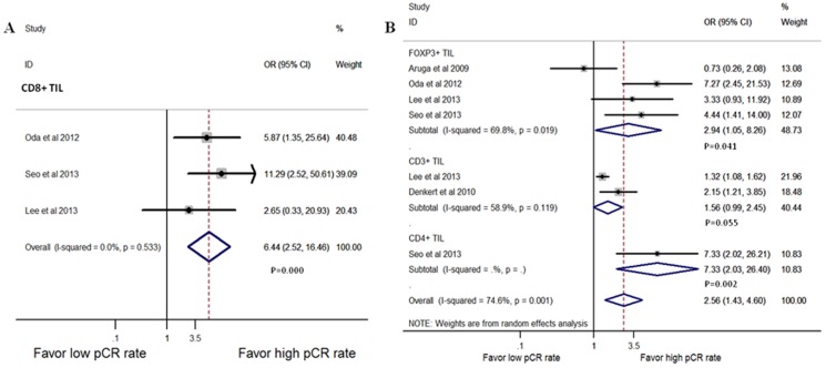
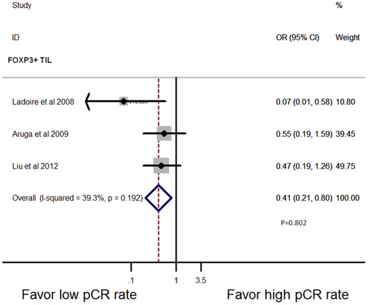
A total of 13 published studies (including 3251 patients) were eligible. In pooled analysis, the detection of higher TILs numbers in pre-treatment biopsy was correlated with better pCR to NAC (OR = 3.93, 95% CI, 3.26-4.73). Moreover, TILs predicted higher pCR rates in triple negative (OR = 2.49, 95% CI: 1.61-3.83), HER2 positive (OR = 5.05, 95% CI: 2.86-8.92) breast cancer, but not in estrogen receptor (ER) positive (OR = 6.21, 95%CI: 0.86-45.15) patients. In multivariate analysis, TILs were still an independent marker for high pCR rate (OR = 1.41, 95% CI: 1.19-1.66). For TILs subset, higher levels of CD8+ and FOXP3+ T-lymphocytes in pre-treatment biopsy respectively predicted better pathological response to NAC (OR = 6.44, 95% CI: 2.52-16.46; OR = 2.94, 95% CI: 1.05-8.26). Only FOXP3+ lymphocytes in post-NAC breast tissue were a predictive marker for low pCR rate in univariate (OR = 0.41, 95% CI: 0.21-0.80) and multivariate (OR = 0.36, 95% CI: 0.13-0.95) analysis.
Higher TILs levels in pre-treatment biopsy indicated higher pCR rates for NAC. TILs subset played different roles in predicting response to NAC.
我们进行了一项系统综述和荟萃分析,以评估肿瘤浸润淋巴细胞(TILs)在乳腺癌新辅助化疗(NAC)反应中的预测作用。
设计了PubMed和Web of Science文献检索。采用随机或固定效应模型估计汇总比值比(OR)。进行异质性和敏感性分析以探索研究间的异质性并评估研究质量的影响。使用漏斗图、Egger检验和Begg检验评估发表偏倚。我们纳入了在乳腺癌NAC中确定TILs和/或TILs亚群对病理完全缓解(pCR)的预测意义的研究。
共有13项已发表研究(包括3251例患者)符合条件。在汇总分析中,治疗前活检中检测到较高的TILs数量与对NAC更好的pCR相关(OR = 3.93,95%CI,3.26 - 4.73)。此外,TILs在三阴性(OR = 2.49,95%CI:1.61 - 3.83)、HER2阳性(OR = 5.05,95%CI:2.86 - 8.92)乳腺癌中预测较高的pCR率,但在雌激素受体(ER)阳性(OR = 6.21,95%CI:0.86 - 45.15)患者中并非如此。在多变量分析中,TILs仍然是高pCR率的独立标志物(OR = 1.41,95%CI:1.19 - 1.66)。对于TILs亚群,治疗前活检中较高水平的CD8 +和FOXP3 + T淋巴细胞分别预测对NAC更好的病理反应(OR = 6.44,95%CI:2.52 - 16.46;OR = 2.94,95%CI:1.05 - 8.26)。仅NAC后乳腺组织中的FOXP3 +淋巴细胞在单变量(OR = 0.41,95%CI:0.21 - 0.80)和多变量(OR = 0.36,95%CI:0.13 - 0.95)分析中是低pCR率的预测标志物。
治疗前活检中较高的TILs水平表明NAC的pCR率较高。TILs亚群在预测对NAC的反应中发挥不同作用。