Guimarães Marcos Duarte, Hochhegger Bruno, Benveniste Marcelo Felipe Kuperman, Odisio Bruno Calazans, Gross Jefferson Luiz, Zurstrassen Charles Edouard, Tyng Chiang Cheng, Bitencourt Almir Galvão Vieira, Marchiori Edson
Department of Imaging, AC Camargo Cancer Center, São Paulo, SP, Brazil.
Department of Radiology, Universidade Federal de Ciências da Saúde de Porto Alegre, Porto Alegre, RS, Brazil.
Clinics (Sao Paulo). 2014 Nov;69(11):787-91. doi: 10.6061/clinics/2014(11)13.
To evaluate the preliminary results obtained using diffusion-weighted magnetic resonance imaging and the apparent diffusion coefficient for planning computed tomography-guided biopsies of selected mediastinal lesions.
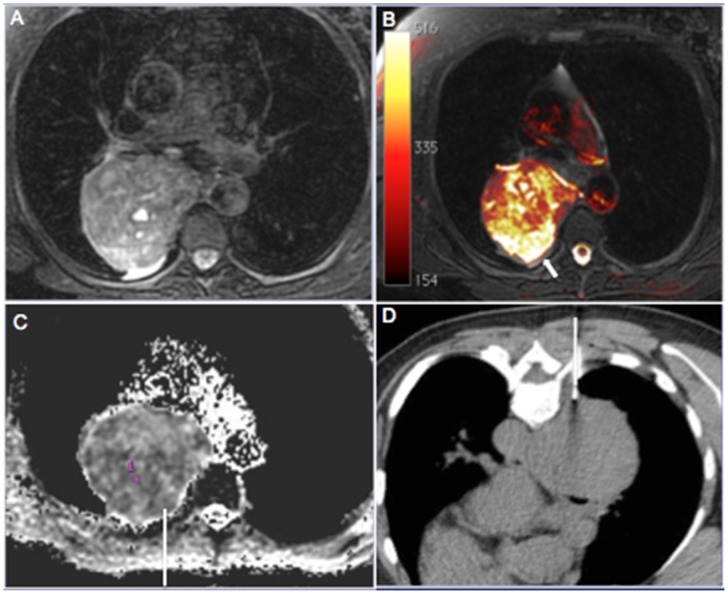
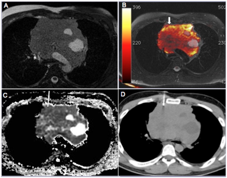
Eight patients with mediastinal lesions suspicious for malignancy were referred for computed tomography-guided biopsy. Diffusion-weighted magnetic resonance imaging and apparent diffusion coefficient measurement were performed to assist in biopsy planning with diffusion/computed tomography fused images. We selected mediastinal lesions that could provide discordant diagnoses depending on the biopsy site, including large heterogeneous masses, lesions associated with lung atelectasis or consolidation, lesions involving large mediastinal vessels and lesions for which the results of biopsy using other methods and histopathological examination were divergent from the clinical and radiological suspicion.
In all cases, the biopsy needle was successfully directed to areas of higher signal intensity on diffusion-weighted sequences and the lowest apparent diffusion coefficient within the lesion (mean, 0.8 [range, 0.6-1.1]×10-3 mm2/s), suggesting high cellularity. All biopsies provided adequate material for specific histopathological diagnoses of four lymphomas, two sarcomas and two thymomas.
Functional imaging tools, such as diffusion-weighted imaging and the apparent diffusion coefficient, are promising for implementation in noninvasive and imaging-guided procedures. However, additional studies are needed to confirm that mediastinal biopsy can be improved with these techniques.
评估使用磁共振扩散加权成像及表观扩散系数来规划对选定纵隔病变进行计算机断层扫描引导活检所获得的初步结果。
8例怀疑为恶性纵隔病变的患者被转诊进行计算机断层扫描引导活检。进行磁共振扩散加权成像及表观扩散系数测量,以利用扩散/计算机断层扫描融合图像辅助活检规划。我们选择了根据活检部位可能得出不一致诊断的纵隔病变,包括大的异质性肿块、与肺不张或实变相关的病变、累及大纵隔血管的病变以及使用其他方法活检结果和组织病理学检查与临床及放射学怀疑不一致的病变。
在所有病例中,活检针均成功指向扩散加权序列上信号强度较高且病变内表观扩散系数最低的区域(平均值为0.8[范围为0.6 - 1.1]×10⁻³mm²/s),提示细胞密度高。所有活检均提供了足够的材料用于对4例淋巴瘤、2例肉瘤和2例胸腺瘤进行特异性组织病理学诊断。
诸如扩散加权成像和表观扩散系数等功能成像工具在无创和成像引导程序中的应用前景广阔。然而,需要进一步研究以证实这些技术可改善纵隔活检。