Sun Wei-Lun, Eisenstein Sarah A, Zelek-Molik Agnieszka, McGinty Jacqueline F
Department of Neurosciences and Neurobiology of Addiction Research Center, Medical University of South Carolina, Charleston, South Carolina, (Drs Sun and McGinty); Department of Psychiatry, Washington University, St Louis, MO (Dr Eisenstein); Department of Brain Biochemistry, Institute of Pharmacology, Polish Academy of Sciences, Krakow, Poland (Dr Zelek-Molik).
Int J Neuropsychopharmacol. 2014 Dec 5;18(1):pyu049. doi: 10.1093/ijnp/pyu049.
Dysregulation in the prefrontal cortex-nucleus accumbens pathway has been implicated in cocaine addiction. We have previously demonstrated that one intra-dorsomedial prefrontal cortex brain-derived neurotrophic factor (BDNF) infusion immediately following the last cocaine self-administration session caused a long-lasting inhibition of cocaine-seeking and normalized the cocaine-induced disturbance of glutamate transmission in the nucleus accumbens after extinction and a cocaine prime. However, the molecular mechanism mediating the brain-derived neurotrophic factor effect on cocaine-induced alterations in extracellular glutamate levels is unknown.
In the present study, we determined the effects of brain-derived neurotrophic factor on cocaine-induced changes in the phosphorylation of synapsin (p-synapsin), a family of presynaptic proteins that mediate synaptic vesicle mobilization, in the nucleus accumbens during early withdrawal.
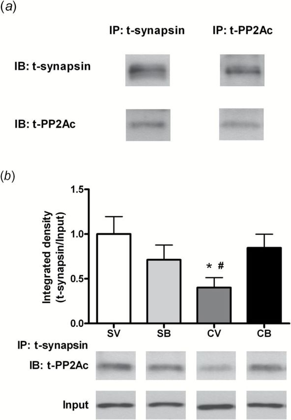
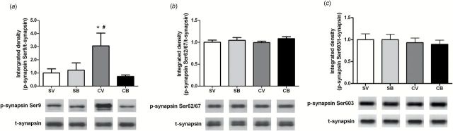
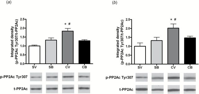
Two hours after cocaine self-administration, p-synapsin Ser9 and p-synapsin Ser62/67, but not p-synapsin Ser603, were increased in the nucleus accumbens. At 22 hours, only p-synapsin Ser9 was still elevated. Elevations at both time points were attenuated by an intra-dorsomedial prefrontal cortex brain-derived neurotrophic factor infusion immediately after the end of cocaine self-administration. Brain-derived neurotrophic factor also reduced cocaine self-administration withdrawal-induced phosphorylation of the protein phosphatase 2A C-subunit, suggesting that brain-derived neurotrophic factor disinhibits protein phosphatase 2A C-subunit, consistent with p-synapsin Ser9 dephosphorylation. Further, co-immunoprecipitation demonstrated that protein phosphatase 2A C-subunit and synapsin are associated in a protein-protein complex that was reduced after 2 hours of withdrawal from cocaine self-administration and reversed by brain-derived neurotrophic factor.
Taken together, these findings demonstrate that brain-derived neurotrophic factor normalizes the cocaine self-administration-induced elevation of p-synapsin in nucleus accumbens that may underlie a disturbance in the probability of neurotransmitter release or represent a compensatory neuroadaptation in response to the hypofunction within the prefrontal cortex-nucleus accumbens pathway during cocaine withdrawal.
前额叶皮质-伏隔核通路的失调与可卡因成瘾有关。我们之前已经证明,在最后一次可卡因自我给药后立即向背内侧前额叶皮质内注射脑源性神经营养因子(BDNF),可长期抑制对可卡因的觅求行为,并使消退和给予可卡因激发后伏隔核中可卡因诱导的谷氨酸传递紊乱恢复正常。然而,介导脑源性神经营养因子对可卡因诱导的细胞外谷氨酸水平变化影响的分子机制尚不清楚。
在本研究中,我们确定了脑源性神经营养因子对可卡因诱导的早期戒断期间伏隔核中突触素(p-突触素)磷酸化变化的影响,突触素是介导突触小泡动员的一类突触前蛋白。
可卡因自我给药两小时后,伏隔核中p-突触素Ser9和p-突触素Ser62/67升高,但p-突触素Ser603未升高。在22小时时,只有p-突触素Ser9仍处于升高状态。可卡因自我给药结束后立即向背内侧前额叶皮质内注射脑源性神经营养因子,可减弱这两个时间点的升高。脑源性神经营养因子还减少了可卡因自我给药戒断诱导的蛋白磷酸酶2A C亚基的磷酸化,表明脑源性神经营养因子使蛋白磷酸酶2A C亚基去抑制,这与p-突触素Ser9的去磷酸化一致。此外,免疫共沉淀表明,蛋白磷酸酶2A C亚基和突触素在一个蛋白质-蛋白质复合物中相互关联,该复合物在可卡因自我给药戒断2小时后减少,并被脑源性神经营养因子逆转。
综上所述,这些发现表明,脑源性神经营养因子使可卡因自我给药诱导的伏隔核中p-突触素升高恢复正常,这可能是神经递质释放概率紊乱的基础,或者代表了可卡因戒断期间前额叶皮质-伏隔核通路功能减退的一种代偿性神经适应。