Volgger Veronika, Sharma Giriraj K, Jing Joseph C, Peaks Ya-Sin A, Loy Anthony Chin, Lazarow Frances, Wang Alex, Qu Yueqiao, Su Erica, Chen Zhongping, Ahuja Gurpreet S, Wong Brian J-F
Department of Otorhinolaryngology-Head and Neck Surgery, Ludwig Maximilian University Munich, 80539 München, Germany.
Department of Otolaryngology-Head and Neck Surgery, University of California Irvine, Orange, CA 92868, USA.
Int J Pediatr Otorhinolaryngol. 2015 Feb;79(2):119-26. doi: 10.1016/j.ijporl.2014.11.019. Epub 2014 Nov 25.
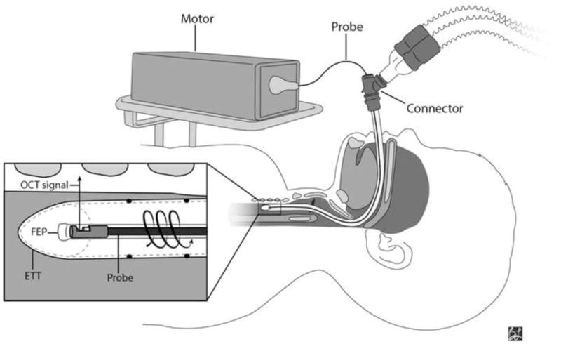
Acquired subglottic stenosis (SGS) most commonly results from prolonged endotracheal intubation and is a diagnostic challenge in the intubated child. At present, no imaging modality allows for in vivo characterization of subglottic microanatomy to identify early signs of acquired SGS while the child remains intubated. Fourier domain optical coherence tomography (FD-OCT) is a minimally invasive, light-based imaging modality which provides high resolution, three dimensional (3D) cross-sectional images of biological tissue. We used long-range FD-OCT to image the subglottis in intubated pediatric patients undergoing minor head and neck surgical procedures in the operating room.
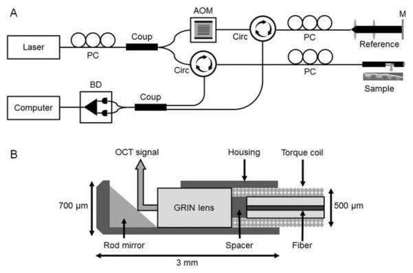
A long-range FD-OCT system and rotary optical probes (1.2mm and 0.7mm outer diameters) were constructed. Forty-six pediatric patients (ages 2-16 years) undergoing minor upper airway surgery (e.g., tonsillectomy and adenoidectomy) were selected for intraoperative, trans-endotracheal tube FD-OCT of the subglottis. Images were analyzed for anatomical landmarks and subepithelial histology. Volumetric image sets were rendered into virtual 3D airway models in Mimics software.
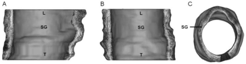
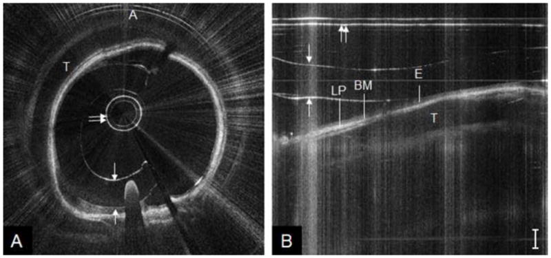
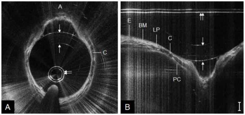
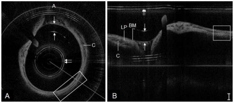
FD-OCT was performed on 46 patients (ages 2-16 years) with no complications. Gross airway contour was visible on all 46 data sets. Twenty (43%) high-quality data sets clearly demonstrated airway anatomy (e.g., tracheal rings, cricoid and vocal folds) and layered microanatomy of the mucosa (e.g., epithelium, basement membrane and lamina propria). The remaining 26 data sets were discarded due to artifact, high signal-to-noise ratio or missing data. 3D airway models were allowed for user-controlled manipulation and multiplanar airway slicing (e.g., sagittal, coronal) for visualization of OCT data at multiple anatomic levels simultaneously.
Long-range FD-OCT produces high-resolution, 3D volumetric images of the pediatric subglottis. This technology offers a safe and practical means for in vivo evaluation of lower airway microanatomy in intubated pediatric patients. Ultimately, FD-OCT may be applied to serial monitoring of the neonatal subglottis in long-term intubated infants at risk for acquired SGS.
获得性声门下狭窄(SGS)最常见于长期气管插管后,对于插管儿童来说是一项诊断挑战。目前,尚无成像方式能够在儿童仍处于插管状态时对声门下微观解剖结构进行活体表征,以识别获得性SGS的早期迹象。傅里叶域光学相干断层扫描(FD-OCT)是一种微创的光学成像方式,可提供生物组织的高分辨率三维(3D)横截面图像。我们使用长程FD-OCT对在手术室接受小型头颈外科手术的插管儿科患者的声门下区域进行成像。
构建了一个长程FD-OCT系统和旋转光学探头(外径分别为1.2mm和0.7mm)。选择46例年龄在2至16岁之间接受小型上呼吸道手术(如扁桃体切除术和腺样体切除术)的儿科患者,在术中通过气管内导管对其声门下区域进行FD-OCT检查。对图像进行解剖标志和上皮下组织学分析。在Mimics软件中将体积图像集渲染为虚拟3D气道模型。
对46例患者(年龄2至16岁)进行了FD-OCT检查,无并发症发生。在所有46个数据集中均可见气道大致轮廓。20个(43%)高质量数据集清晰显示了气道解剖结构(如气管环、环状软骨和声带)以及黏膜的分层微观解剖结构(如上皮、基底膜和固有层)。其余26个数据集因伪影、高信噪比或数据缺失而被丢弃。3D气道模型允许用户进行控制操作和多平面气道切片(如矢状面、冠状面),以便同时在多个解剖层面可视化OCT数据。
长程FD-OCT可生成儿科声门下区域的高分辨率3D体积图像。该技术为活体评估插管儿科患者的下呼吸道微观解剖结构提供了一种安全实用的方法。最终,FD-OCT可能应用于对有获得性SGS风险的长期插管婴儿的新生儿声门下区域进行连续监测。