Yang Song-ran, Shang Xin-yuan, Tao Jun, Liu Jian-yang, Hua Ping
Department of Experimental Psychology, University of Oxford, Oxford, United Kingdom.
Department of Neurology, Guangzhou First People's Hospital, Guangzhou Medical University, Guangzhou, China.
PLoS One. 2015 Jan 2;10(1):e116168. doi: 10.1371/journal.pone.0116168. eCollection 2015.
To explore the structural basis of post-stroke apathy by using voxel-based analysis (VBA) of fractional anisotropy (FA) maps.
We enrolled 54 consecutive patients with ischemic stroke during convalescence, and divided them into apathy (n = 31) and non-apathy (n = 23) groups. We obtained magnetic resonance images of their brains, including T1, T2 and DTI sequences. Age, sex, education level, Hamilton Depression Scale (HAMD) scores, Mini-Mental State Examination (MMSE) scores, National Institutes of Health Stroke Scale (NIHSS) scores, and infarct locations for the two groups were compared. Finally, to investigate the structural basis of post-stroke apathy, VBA of FA maps was performed in which we included the variables that a univariate analysis determined had P-values less than 0.20 as covariates.
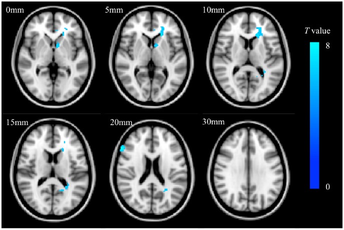
HAMD (P = 0.01) and MMSE (P<0.01) scores differed significantly between the apathy and non-apathy groups. After controlling for age, education level, HAMD scores, and MMSE scores, significant FA reduction was detected in four clusters with peak voxels at the genu of the corpus callosum (X = -16, Y = 30, Z = 8), left anterior corona radiata (-22, 30, 10), splenium of the corpus callosum (-24, -56, 18), and right inferior frontal gyrus white matter (52, 24, 18), after family-wise error correction for multiple comparisons.
Post-stroke apathy is related to depression and cognitive decline. Damage to the genu of the corpus callosum, left anterior corona radiata, splenium of the corpus callosum, and white matter in the right inferior frontal gyrus may lead to apathy after ischemic stroke.
通过基于体素的分数各向异性(FA)图分析(VBA)探讨中风后冷漠的结构基础。
我们连续纳入了54例恢复期缺血性中风患者,并将他们分为冷漠组(n = 31)和非冷漠组(n = 23)。我们获取了他们大脑的磁共振图像,包括T1、T2和DTI序列。比较了两组的年龄、性别、教育水平、汉密尔顿抑郁量表(HAMD)评分、简易精神状态检查表(MMSE)评分、美国国立卫生研究院卒中量表(NIHSS)评分以及梗死部位。最后,为了研究中风后冷漠的结构基础,我们进行了FA图的VBA分析,其中我们将单变量分析确定的P值小于0.20的变量作为协变量纳入。
冷漠组和非冷漠组之间的HAMD(P = 0.01)和MMSE(P<0.01)评分存在显著差异。在控制年龄、教育水平、HAMD评分和MMSE评分后,在胼胝体膝部(X = -16,Y = 30,Z = 8)、左侧放射冠前部(-22,30,10)、胼胝体压部(-24,-56,18)和右侧额下回白质(52,24,18)的四个簇中检测到显著的FA降低,这是在对多次比较进行家族性错误校正之后。
中风后冷漠与抑郁和认知下降有关。胼胝体膝部、左侧放射冠前部、胼胝体压部和右侧额下回白质的损伤可能导致缺血性中风后出现冷漠。