Chazeau Anaël, Garcia Mikael, Czöndör Katalin, Perrais David, Tessier Béatrice, Giannone Grégory, Thoumine Olivier
Interdisciplinary Institute for Neuroscience, University of Bordeaux, Unité Mixte de Recherche 5297, F-33000 Bordeaux, France Interdisciplinary Institute for Neuroscience, Centre Nationale de la Recherche Scientifique, Unité Mixte de Recherche 5297, F-33000 Bordeaux, France.
Interdisciplinary Institute for Neuroscience, University of Bordeaux, Unité Mixte de Recherche 5297, F-33000 Bordeaux, France Interdisciplinary Institute for Neuroscience, Centre Nationale de la Recherche Scientifique, Unité Mixte de Recherche 5297, F-33000 Bordeaux, France CYTOO, Minatec, Grenoble, 38054 Grenoble, France.
Mol Biol Cell. 2015 Mar 1;26(5):859-73. doi: 10.1091/mbc.E14-06-1086. Epub 2015 Jan 7.
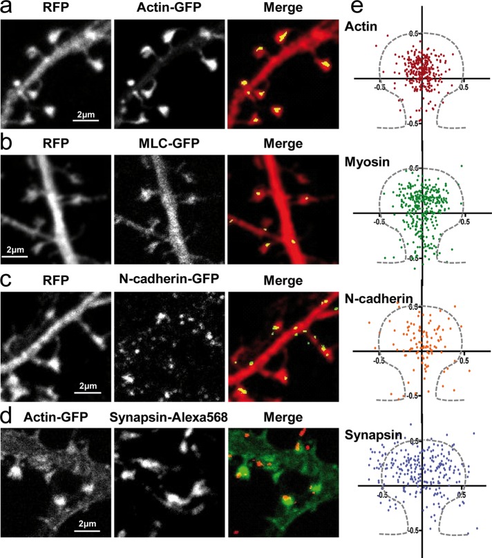
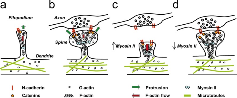
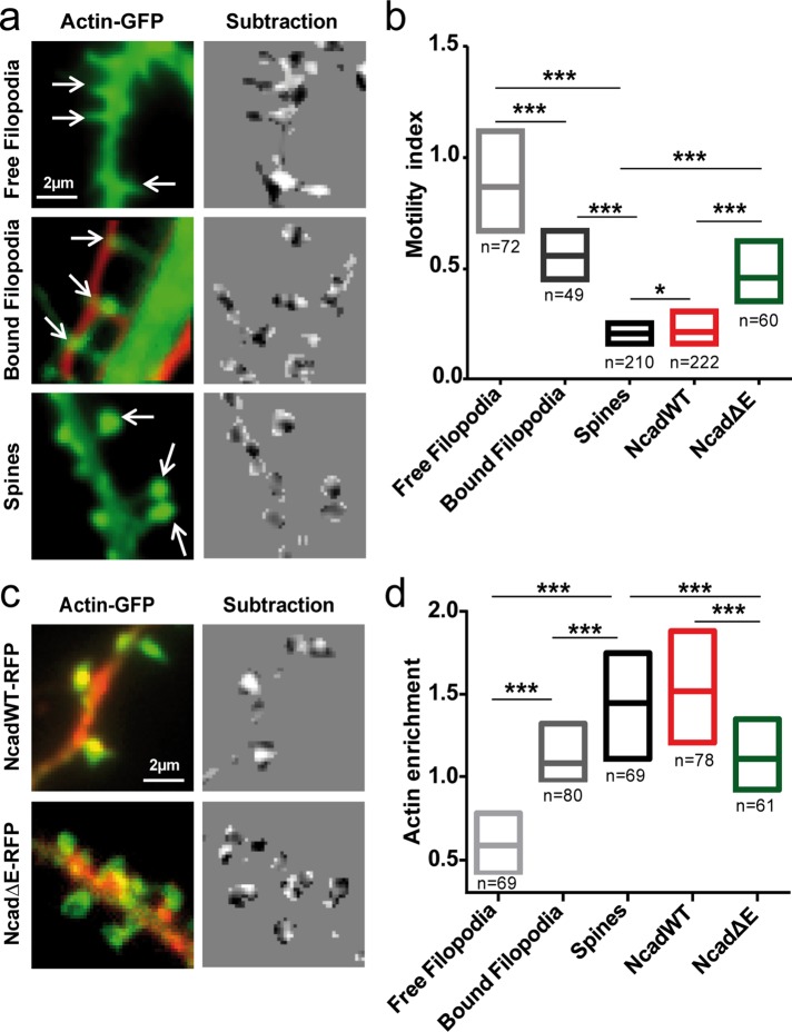
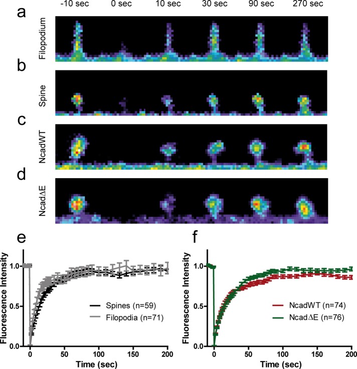
The morphology of neuronal dendritic spines is a critical indicator of synaptic function. It is regulated by several factors, including the intracellular actin/myosin cytoskeleton and transcellular N-cadherin adhesions. To examine the mechanical relationship between these molecular components, we performed quantitative live-imaging experiments in primary hippocampal neurons. We found that actin turnover and structural motility were lower in dendritic spines than in immature filopodia and increased upon expression of a nonadhesive N-cadherin mutant, resulting in an inverse relationship between spine motility and actin enrichment. Furthermore, the pharmacological stimulation of myosin II induced the rearward motion of actin structures in spines, showing that myosin II exerts tension on the actin network. Strikingly, the formation of stable, spine-like structures enriched in actin was induced at contacts between dendritic filopodia and N-cadherin-coated beads or micropatterns. Finally, computer simulations of actin dynamics mimicked various experimental conditions, pointing to the actin flow rate as an important parameter controlling actin enrichment in dendritic spines. Together these data demonstrate that a clutch-like mechanism between N-cadherin adhesions and the actin flow underlies the stabilization of dendritic filopodia into mature spines, a mechanism that may have important implications in synapse initiation, maturation, and plasticity in the developing brain.
神经元树突棘的形态是突触功能的关键指标。它受多种因素调节,包括细胞内肌动蛋白/肌球蛋白细胞骨架和跨细胞N-钙黏蛋白黏附。为了研究这些分子成分之间的力学关系,我们在原代海马神经元中进行了定量实时成像实验。我们发现,树突棘中的肌动蛋白周转和结构运动性低于未成熟丝状伪足,并且在表达非黏附性N-钙黏蛋白突变体时增加,导致棘突运动性与肌动蛋白富集之间呈负相关。此外,对肌球蛋白II的药理学刺激诱导了棘突中肌动蛋白结构的向后运动,表明肌球蛋白II对肌动蛋白网络施加张力。令人惊讶的是,在树突丝状伪足与N-钙黏蛋白包被的珠子或微图案之间的接触处诱导形成了富含肌动蛋白的稳定的、类似棘突的结构。最后,肌动蛋白动力学的计算机模拟模拟了各种实验条件,指出肌动蛋白流速是控制树突棘中肌动蛋白富集的一个重要参数。这些数据共同表明,N-钙黏蛋白黏附与肌动蛋白流之间的类似离合器的机制是树突丝状伪足稳定为成熟棘突的基础,这一机制可能对发育中大脑的突触起始、成熟和可塑性具有重要意义。