Wang Huaijun, Kaneko Osamu F, Tian Lu, Hristov Dimitre, Willmann Jürgen K
From the Departments of *Radiology, Molecular Imaging Program at Stanford, School of Medicine, †Health, Research and Policy, and ‡Radiation Oncology, Stanford University, Stanford, CA.
Invest Radiol. 2015 May;50(5):322-9. doi: 10.1097/RLI.0000000000000128.
We sought to assess the feasibility and reproducibility of 3-dimensional ultrasound molecular imaging (USMI) of vascular endothelial growth factor receptor 2 (VEGFR2) expression in tumor angiogenesis using a clinical matrix array transducer and a clinical grade VEGFR2-targeted contrast agent in a murine model of human colon cancer.
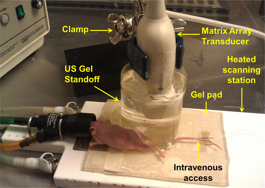
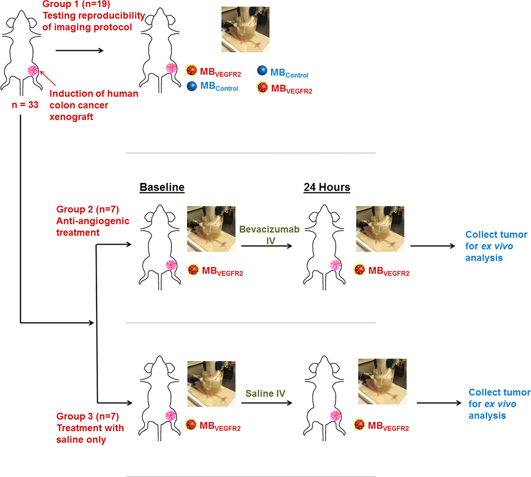
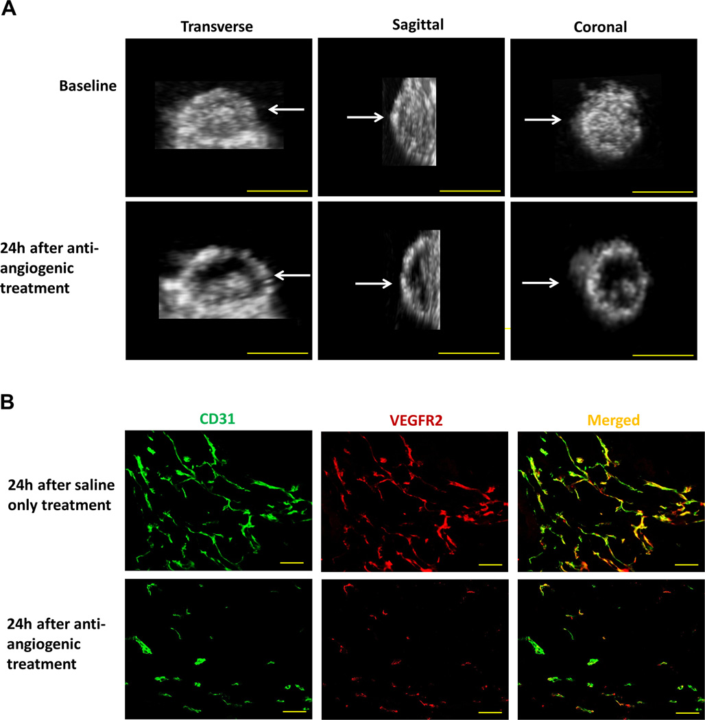
Animal studies were approved by the Institutional Administrative Panel on Laboratory Animal Care. Mice with human colon cancer xenografts (n = 33) were imaged with a clinical ultrasound system and transducer (Philips iU22; X6-1) after intravenous injection of either clinical grade VEGFR2-targeted microbubbles or nontargeted control microbubbles. Nineteen mice were scanned twice to assess imaging reproducibility. Fourteen mice were scanned both before and 24 hours after treatment with either bevacizumab (n = 7) or saline only (n = 7). Three-dimensional USMI data sets were retrospectively reconstructed into multiple consecutive 1-mm-thick USMI data sets to simulate 2-dimensional imaging. Vascular VEGFR2 expression was assessed ex vivo using immunofluorescence.
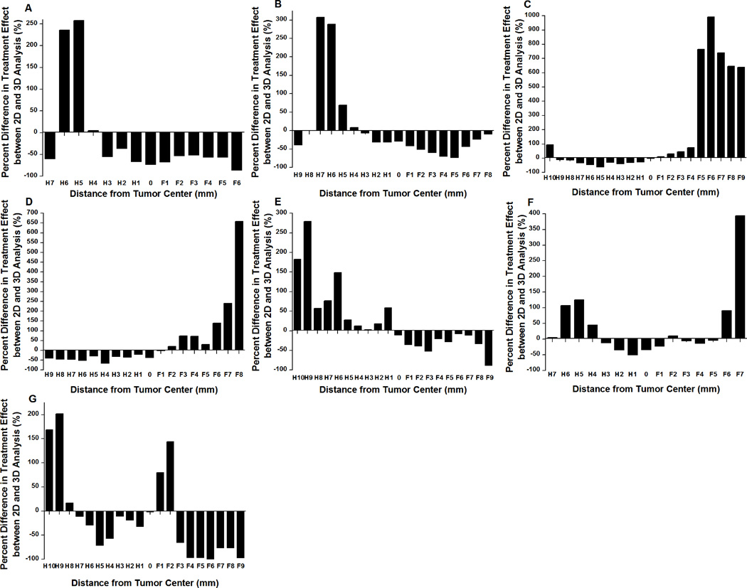
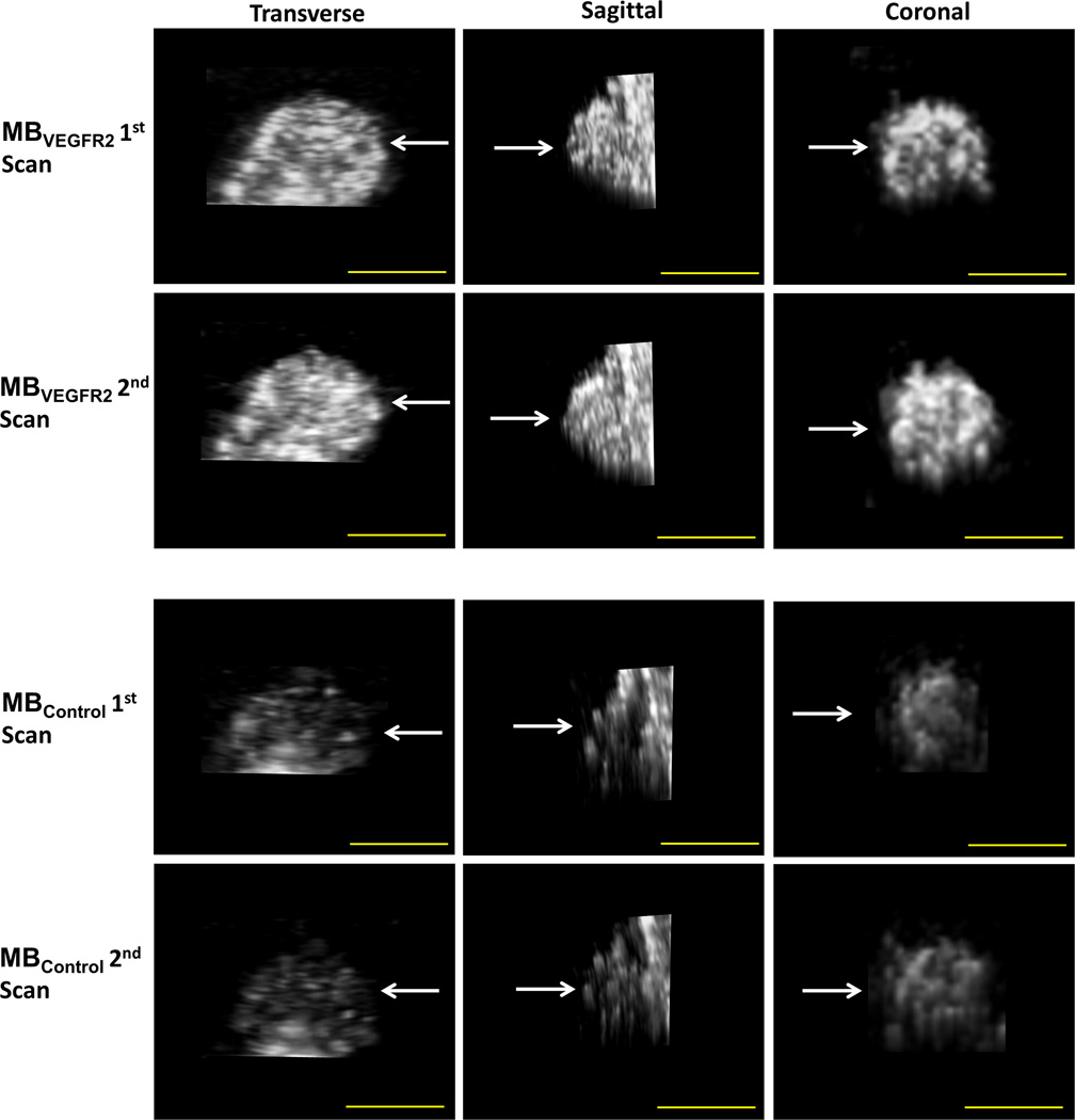
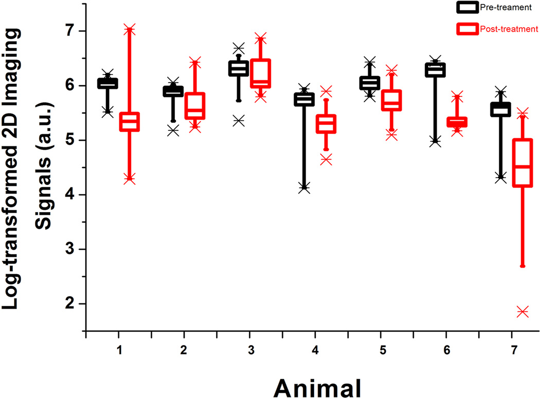
Three-dimensional USMI was highly reproducible using both VEGFR2-targeted microbubbles and nontargeted control microbubbles (intraclass correlation coefficient, 0.83). The VEGFR2-targeted USMI signal significantly (P = 0.02) decreased by 57% after antiangiogenic treatment compared with the control group, which correlated well with ex vivo VEGFR2 expression on immunofluorescence (ρ = 0.93, P = 0.003). If only central 1-mm tumor planes were analyzed to assess antiangiogenic treatment response, the USMI signal change was significantly (P = 0.006) overestimated by an average of 27% (range, 2%-73%) compared with 3-dimensional USMI.
Three-dimensional USMI is feasible and highly reproducible and allows accurate assessment and monitoring of VEGFR2 expression in tumor angiogenesis in a murine model of human colon cancer.
我们试图使用临床矩阵阵列换能器和临床级血管内皮生长因子受体2(VEGFR2)靶向造影剂,在人结肠癌小鼠模型中评估三维超声分子成像(USMI)检测肿瘤血管生成中VEGFR2表达的可行性和可重复性。
动物研究经机构实验动物护理管理小组批准。将人结肠癌异种移植小鼠(n = 33)静脉注射临床级VEGFR2靶向微泡或非靶向对照微泡后,用临床超声系统和换能器(飞利浦iU22;X6 - 1)进行成像。19只小鼠进行了两次扫描以评估成像的可重复性。14只小鼠在使用贝伐单抗(n = 7)或仅用生理盐水(n = 7)治疗前和治疗后24小时进行扫描。三维USMI数据集被回顾性重建为多个连续的1毫米厚的USMI数据集,以模拟二维成像。使用免疫荧光在体外评估血管VEGFR2表达。
使用VEGFR2靶向微泡和非靶向对照微泡时,三维USMI具有高度可重复性(组内相关系数,0.83)。与对照组相比,抗血管生成治疗后,VEGFR2靶向USMI信号显著(P = 0.02)降低了57%,这与免疫荧光体外VEGFR2表达密切相关(ρ = 0.93,P = 0.003)。如果仅分析中央1毫米肿瘤平面来评估抗血管生成治疗反应,与三维USMI相比,USMI信号变化被显著(P = 0.006)高估,平均高估27%(范围,2% - 73%)。
三维USMI是可行的且具有高度可重复性,能够准确评估和监测人结肠癌小鼠模型中肿瘤血管生成中的VEGFR2表达。