Abou-Elkacem Lotfi, Wilson Katheryne E, Johnson Sadie M, Chowdhury Sayan M, Bachawal Sunitha, Hackel Benjamin J, Tian Lu, Willmann Jürgen K
1. Department of Radiology, Molecular Imaging Program at Stanford, Stanford University, School of Medicine, Stanford, California, USA;
2. Department of Chemical Engineering and Materials Science, University of Minnesota, Minnesota, USA;
Theranostics. 2016 Jun 28;6(11):1740-52. doi: 10.7150/thno.15169. eCollection 2016.
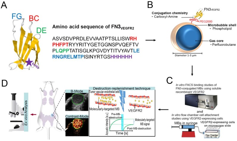
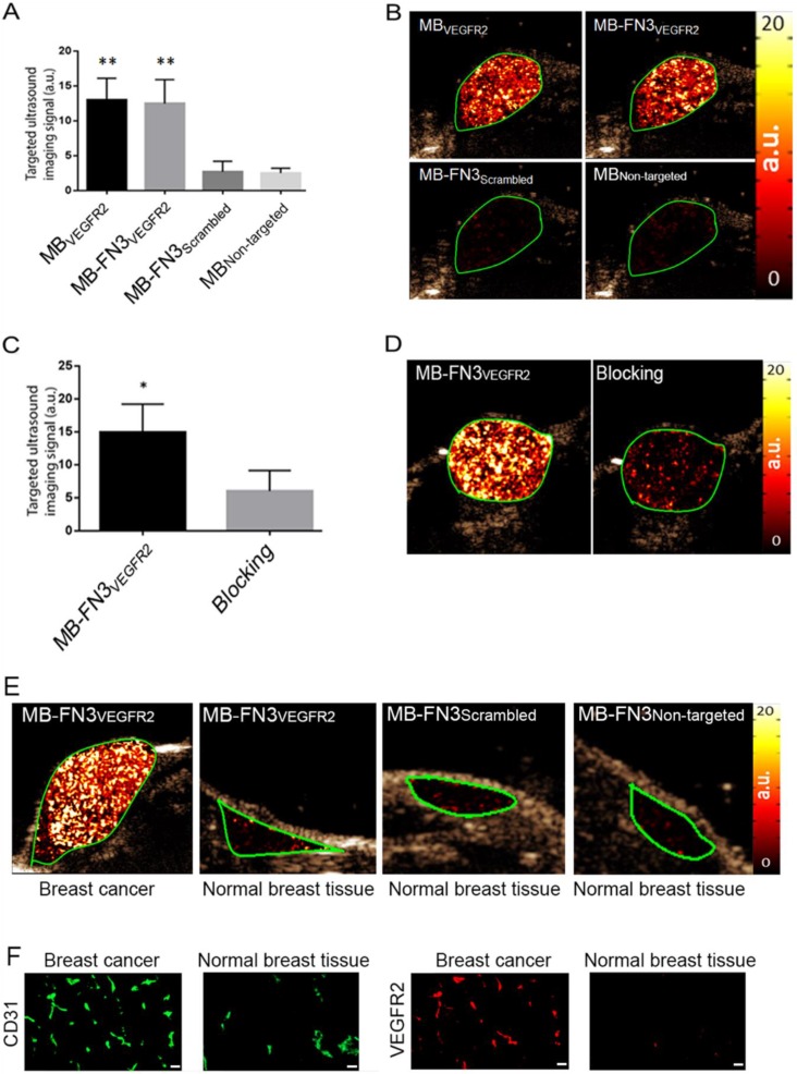
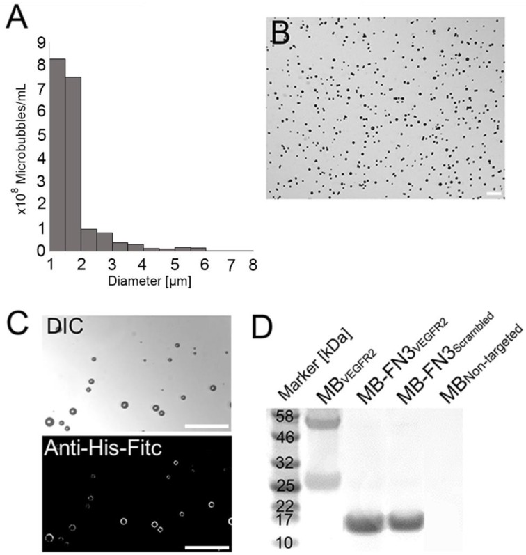
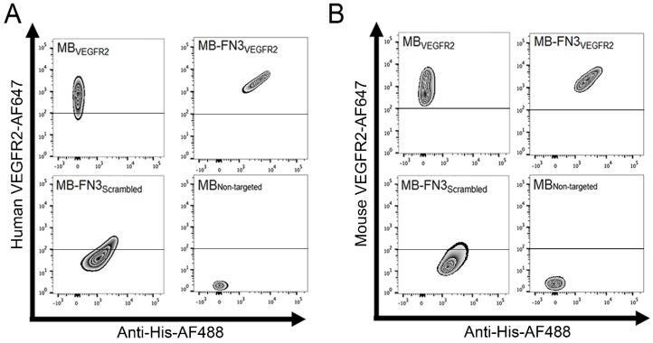
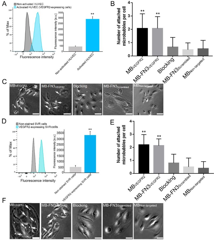
Molecularly-targeted microbubbles (MBs) are increasingly being recognized as promising contrast agents for oncological molecular imaging with ultrasound. With the detection and validation of new molecular imaging targets, novel binding ligands are needed that bind to molecular imaging targets with high affinity and specificity. In this study we assessed a novel class of potentially clinically translatable MBs using an engineered 10(th) type III domain of human-fibronectin (MB-FN3VEGFR2) scaffold-ligand to image VEGFR2 on the neovasculature of cancer. The in vitro binding of MB-FN3VEGFR2 to a soluble VEGFR2 was assessed by flow-cytometry (FACS) and binding to VEGFR2-expressing cells was assessed by flow-chamber cell attachment studies under flow shear stress conditions. In vivo binding of MB-FN3VEGFR2 was tested in a transgenic mouse model (FVB/N Tg(MMTV/PyMT634Mul) of breast cancer and control litter mates with normal mammary glands. In vitro FACS and flow-chamber cell attachment studies showed significantly (P<0.01) higher binding to VEGFR2 using MB-FN3VEGFR2 than control agents. In vivo ultrasound molecular imaging (USMI) studies using MB-FN3VEGFR2 demonstrated specific binding to VEGFR2 and was significantly higher (P<0.01) in breast cancer compared to normal breast tissue. Ex vivo immunofluorescence-analysis showed significantly (P<0.01) increased VEGFR2-expression in breast cancer compared to normal mammary tissue. Our results suggest that MBs coupled to FN3-scaffolds can be designed and used for USMI of breast cancer neoangiogenesis. Due to their small size, stability, solubility, the lack of glycosylation and disulfide bonds, FN3-scaffolds can be recombinantly produced with the advantage of generating small, high affinity ligands in a cost efficient way for USMI.
分子靶向微泡(MBs)越来越被认为是用于超声肿瘤分子成像的有前景的造影剂。随着新的分子成像靶点的检测和验证,需要能够以高亲和力和特异性结合分子成像靶点的新型结合配体。在本研究中,我们使用人纤连蛋白工程化的第10个III型结构域(MB-FN3VEGFR2)支架-配体评估了一类新型的、具有潜在临床可转化性的微泡,以对癌症新生血管上的VEGFR2进行成像。通过流式细胞术(FACS)评估MB-FN3VEGFR2与可溶性VEGFR2的体外结合,并在流动剪切应力条件下通过流动腔细胞附着研究评估其与表达VEGFR2的细胞的结合。在乳腺癌转基因小鼠模型(FVB/N Tg(MMTV/PyMT634Mul))和具有正常乳腺的对照同窝仔鼠中测试了MB-FN3VEGFR2的体内结合。体外FACS和流动腔细胞附着研究表明,与对照剂相比,使用MB-FN3VEGFR2时与VEGFR2的结合显著更高(P<0.01)。使用MB-FN3VEGFR2的体内超声分子成像(USMI)研究表明其与VEGFR2特异性结合,并且与正常乳腺组织相比,在乳腺癌中显著更高(P<0.01)。离体免疫荧光分析表明,与正常乳腺组织相比,乳腺癌中VEGFR2表达显著增加(P<0.01)。我们的结果表明,与FN3支架偶联的微泡可被设计并用于乳腺癌新生血管生成的超声分子成像。由于其尺寸小、稳定性好、溶解性好、缺乏糖基化和二硫键,FN3支架可以重组生产,具有以经济高效的方式为超声分子成像生成小的、高亲和力配体的优势。