Shirkavand Saeed, Moslehifard Elnaz
Assistant Professor, Department of Prosthodontics, Faculty of Dentistry, Urmia University of Medical Sciences, Urmia, Iran.
Dental and Periodontal Research Center, Tabriz University of Medical Sciences, Tabriz, Iran ; Assistant Professor, Department of Prosthodontics, Faculty of Dentistry, Tabriz University of Medical Sciences, Tabriz, Iran.
J Dent Res Dent Clin Dent Prospects. 2014 Fall;8(4):197-203. doi: 10.5681/joddd.2014.036. Epub 2014 Dec 3.
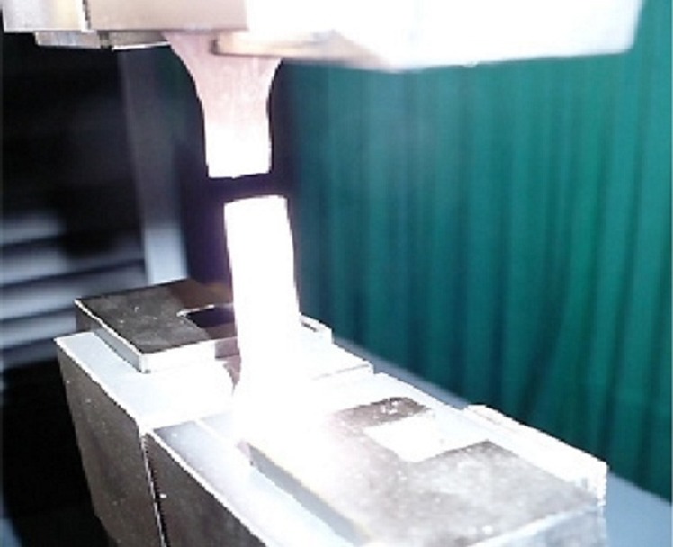
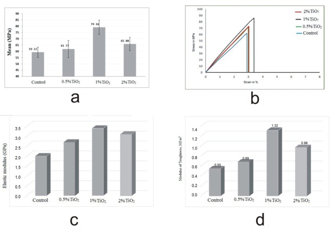
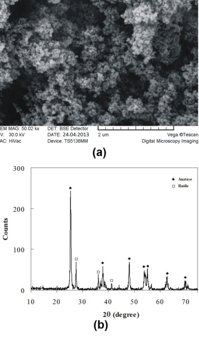
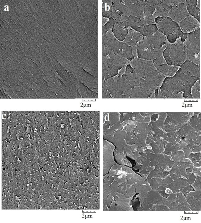
Background and aims. Adding further fillers to dental resins may enhance their physical characteristics. The aim of this study was to evaluate the tensile strength of heat-curing acrylic resin reinforced by TiO2nanoparticles added into the resin matrix. Materials and methods. Commercially available TiO2 nanoparticles were obtained and characterized using X-ray diffrac-tion (XRD) and scanning electron microscopy (SEM) to determine their crystalline structure, particle size and morphology. TiO2-acrylic resin nanocomposite was prepared by mixing 0.5, 1 and 2 (wt%) of surface modified TiO2 nanoparticles in an amalgamator providing three groups of samples. Before curing, the obtained paste was packed into steel molds. After cur-ing, the specimens were removed from the molds. The tensile strength test samples were prepared according to ISO 1567. Results. Two crystalline phases were found in TiO2 nanoparticles including: (i) anatase as the major one, and (ii) rutile. The average particle size calculated according to the Scherrer equation was 20.4 nm, showing a normal size distribution. According to SEM images, the nanocomposite with 1wt% TiO2 nanoparticles had a better distribution compared to other groups. In addition, the group by 1wt% TiO2 exhibited higher tensile strength with a significant difference compared to other groups. ANOVA showed significant differences between the contents of TiO2 particles in acrylic resin (F = 22.19; P < 0.001). Conclusion. A considerable increase in tensile strength was observed with titania NPs reinforcement agents in 1wt% by weight. Further increase of TiO2 nanoparticles decreased the tensile strength.
背景与目的。向牙科树脂中添加更多填料可能会增强其物理特性。本研究的目的是评估添加到树脂基体中的二氧化钛纳米颗粒增强热固化丙烯酸树脂的拉伸强度。材料与方法。获取市售的二氧化钛纳米颗粒,并使用X射线衍射(XRD)和扫描电子显微镜(SEM)对其进行表征,以确定其晶体结构、粒径和形态。通过在混汞器中混合0.5%、1%和2%(重量)的表面改性二氧化钛纳米颗粒制备二氧化钛-丙烯酸树脂纳米复合材料,从而得到三组样品。固化前,将所得糊剂装入钢模具中。固化后,将试样从模具中取出。根据ISO 1567制备拉伸强度测试样品。结果。在二氧化钛纳米颗粒中发现了两个晶相,包括:(i)以锐钛矿为主相,以及(ii)金红石相。根据谢乐方程计算的平均粒径为20.4 nm,呈现正态尺寸分布。根据SEM图像,与其他组相比,含有1%重量二氧化钛纳米颗粒的纳米复合材料具有更好的分布。此外,含有1%重量二氧化钛的组表现出更高的拉伸强度,与其他组相比有显著差异。方差分析表明丙烯酸树脂中二氧化钛颗粒含量之间存在显著差异(F = 22.19;P < 0.001)。结论。观察到用1%重量的二氧化钛纳米颗粒增强剂可使拉伸强度显著提高。二氧化钛纳米颗粒含量的进一步增加会降低拉伸强度。