Division of Biomedical Cell Biology, Warwick Medical School, Gibbet Hill Road, Coventry, CV4 7AL, UK.
Cancer Research UK Leicester Centre and Department of Biochemistry, University of Leicester, Leicester LE1 9HN, UK.
Biol Open. 2015 Jan 16;4(2):170-9. doi: 10.1242/bio.201410843.
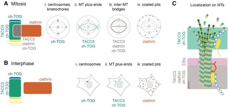
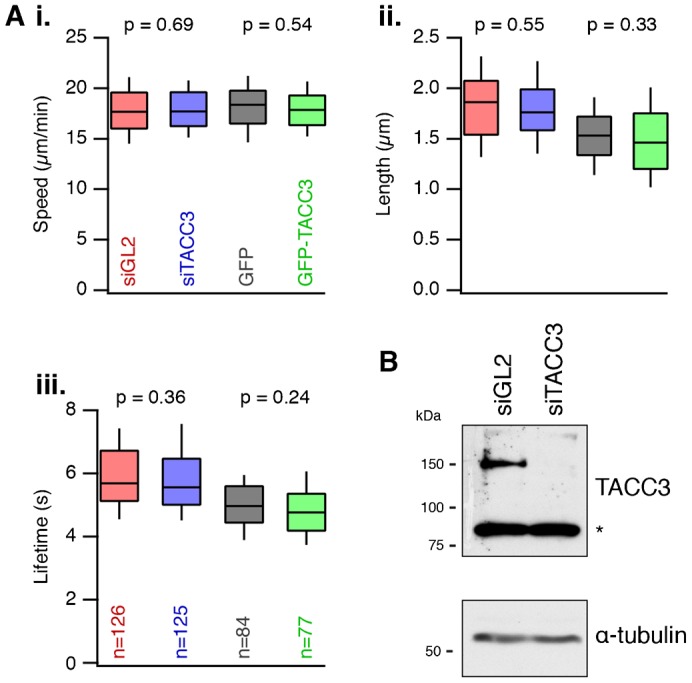
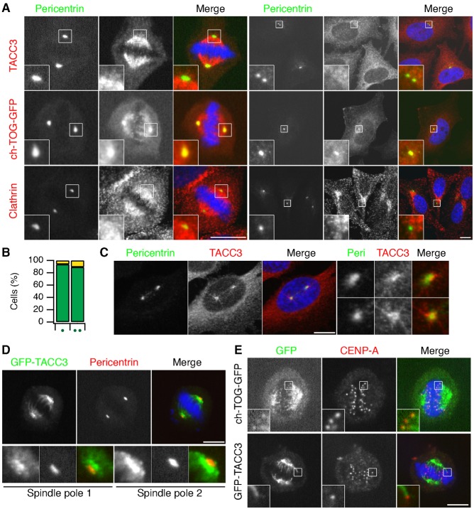
The interaction between TACC3 (transforming acidic coiled coil protein 3) and the microtubule polymerase ch-TOG (colonic, hepatic tumor overexpressed gene) is evolutionarily conserved. Loading of TACC3-ch-TOG onto mitotic spindle microtubules requires the phosphorylation of TACC3 by Aurora-A kinase and the subsequent interaction of TACC3 with clathrin to form a microtubule-binding surface. Recent work indicates that TACC3 can track the plus-ends of microtubules and modulate microtubule dynamics in non-dividing cells via its interaction with ch-TOG. Whether there is a pool of TACC3-ch-TOG that is independent of clathrin in human cells, and what is the function of this pool, are open questions. Here, we describe the molecular interaction between TACC3 and ch-TOG that permits TACC3 recruitment to the plus-ends of microtubules. This TACC3-ch-TOG pool is independent of EB1, EB3, Aurora-A phosphorylation and binding to clathrin. We also describe the distinct combinatorial subcellular pools of TACC3, ch-TOG and clathrin. TACC3 is often described as a centrosomal protein, but we show that there is no significant population of TACC3 at centrosomes. The delineation of distinct protein pools reveals a simplified view of how these proteins are organized and controlled by post-translational modification.
TACC3(转化酸性卷曲螺旋蛋白 3)与微管聚合酶 ch-TOG(结肠、肝肿瘤过表达基因)之间的相互作用在进化上是保守的。TACC3-ch-TOG 加载到有丝分裂纺锤体微管上需要 Aurora-A 激酶对 TACC3 的磷酸化,以及随后 TACC3 与网格蛋白的相互作用,以形成微管结合表面。最近的工作表明,TACC3 可以通过与 ch-TOG 的相互作用追踪微管的正极,并调节非分裂细胞中的微管动力学。在人类细胞中是否存在独立于网格蛋白的 TACC3-ch-TOG 池,以及该池的功能是什么,这些都是悬而未决的问题。在这里,我们描述了 TACC3 和 ch-TOG 之间的分子相互作用,允许 TACC3 招募到微管的正极。这个 TACC3-ch-TOG 池独立于 EB1、EB3、Aurora-A 磷酸化和与网格蛋白的结合。我们还描述了 TACC3、ch-TOG 和网格蛋白的不同组合亚细胞池。TACC3 通常被描述为中心体蛋白,但我们表明中心体中没有大量的 TACC3。不同蛋白质池的描绘揭示了这些蛋白质如何通过翻译后修饰进行组织和控制的简化视图。