Vincenzi Matteo, Pasquotti Giulio, Polverosi Roberta, Pasquali Claudio, Pomerri Fabio
Cancer Imaging. 2014 Apr 22;14(1):5. doi: 10.1186/1470-7330-14-5.
To describe the main imaging characteristics of pancreatic metastases from renal cell carcinoma (RCC) with particular attention to CT features, underlining possible criteria for a differential diagnosis.
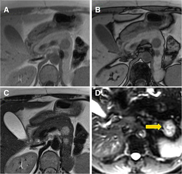
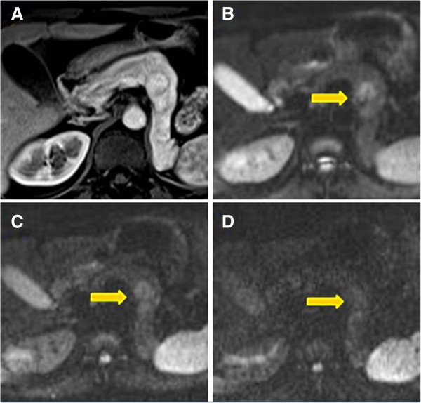
15 patients have been included in this study. 14 patients underwent multislice CT with triphasic acquisition (unenhanced, pancreatic parenchymal and portal venous phases). In 9 cases a delayed phase (120 sec) was also acquired. 5 patients underwent MRI, before and after administration of gadolinium.
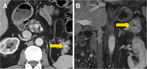
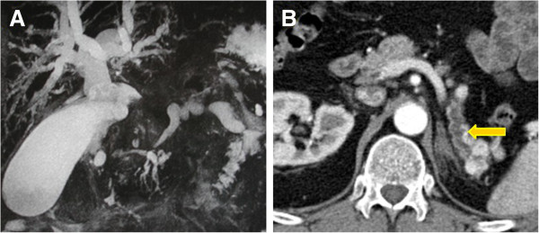
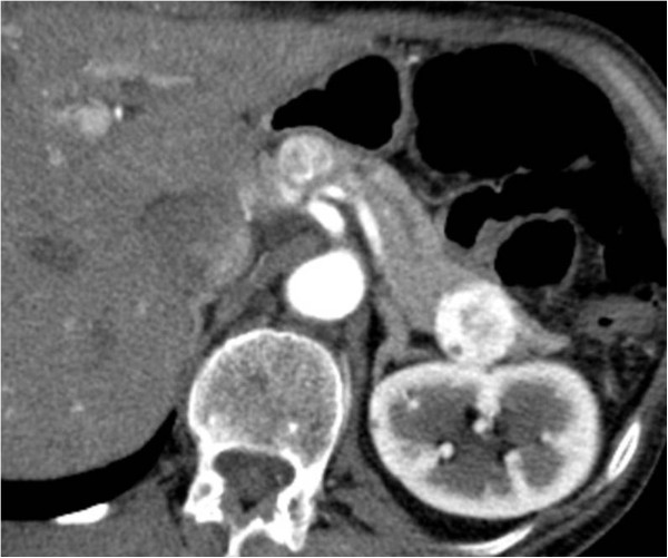
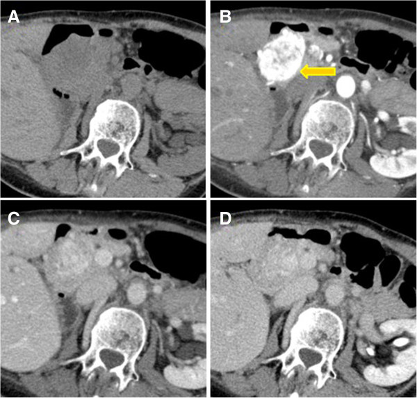
The mean time interval between nephrectomy and recurrence was 7.5 years (range 1-17 years). On CT metastases avidly enhanced in the parenchymal phase and then demonstrated a significant wash-out, approaching isodensity to the normal pancreatic parenchyma in the portal phase. In the portal phase 20 of the 25 lesions found in the arterial phase were recognizable. On non-enhanced scans, only 13 of the 25 lesions were detected.
Renal Cell Carcinomas require a prolonged CT or MRI follow-up.
描述肾细胞癌(RCC)胰腺转移灶的主要影像学特征,特别关注CT表现,强调可能的鉴别诊断标准。
本研究纳入15例患者。14例患者接受了多排螺旋CT三相扫描(平扫、胰腺实质期和门静脉期)。9例还进行了延迟期(120秒)扫描。5例患者在注射钆对比剂前后接受了MRI检查。
肾切除与复发之间的平均时间间隔为7.5年(范围1 - 17年)。CT上,转移灶在实质期明显强化,随后显著廓清,在门静脉期接近正常胰腺实质的等密度。在门静脉期,动脉期发现的25个病灶中有20个可识别。在平扫上,25个病灶中仅检测到13个。
肾细胞癌需要进行长时间的CT或MRI随访。