Kaihola Helena, Olivier Jocelien, Poromaa Inger Sundström, Åkerud Helena
Department of Women's and Children's Health, Uppsala University, Uppsala, Sweden.
Department of Women's and Children's Health, Uppsala University, Uppsala, Sweden; Department of Behavioural Physiology, University of Groningen, Groningen, The Netherlands.
PLoS One. 2015 Jan 22;10(1):e0116459. doi: 10.1371/journal.pone.0116459. eCollection 2015.
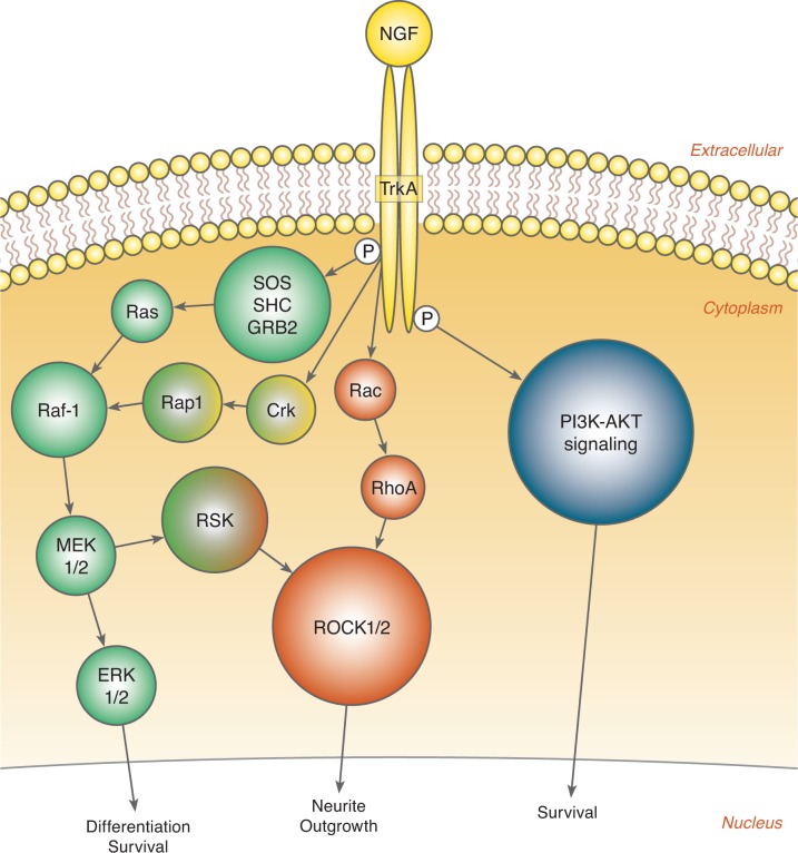
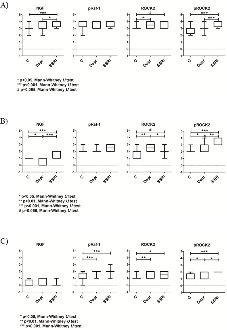
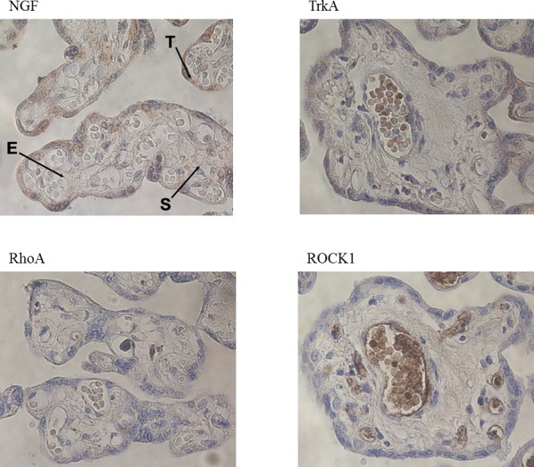
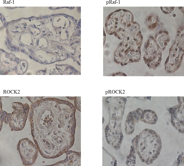
Depressive symptoms during pregnancy are common and may have impact on the developing child. Selective serotonin reuptake inhibitors (SSRIs) are the most prescribed antidepressant treatment, but unfortunately, these treatments can also negatively affect the behavioral development and health of a child during pregnancy. In addition, serotonin (5-HT) exerts neurotrophic actions with thus far not fully known effects in the offspring. The neurotrophic growth factor (NGF) is involved in neuronal cell survival and differentiation, and altered placenta levels have been found to increase the risk for pregnancy complications, similar to those found in women treated with SSRIs. We therefore investigated whether the NGF signaling pathway was altered in the placenta from women treated with SSRIs (n = 12) and compared them with placenta from depressed (n = 12) and healthy mothers (n = 12). Results from immunohistochemical stainings revealed that placental NGF protein levels of SSRI-treated women were increased in both trophoblasts and endothelial cells compared with depressed and control women. In addition, downstream of the NGF receptor TrkA, increased levels of the signaling proteins ROCK2 and phosphorylated Raf-1 were found in stromal cells and a tendency towards increased levels of ROCK2 in trophoblasts and endothelial cells in SSRI-treated women when compared to healthy controls. SSRI-treated women also displayed increased levels of phosphorylated ROCK2 in all placental cell types studied in comparison with depressed and control women. Interestingly, in placental endothelial cells from depressed women, NGF levels were significantly lower compared to control women, but ROCK2 levels were increased compared with control and SSRI-treated women. Taken together, these results show that the NGF signaling and downstream pathways in the placenta are affected by SSRI treatment and/or antenatal depression. This might lead to an altered placental function, although the clinical relevance of our findings still needs to be investigated.
孕期抑郁症状很常见,且可能会对发育中的胎儿产生影响。选择性5-羟色胺再摄取抑制剂(SSRI)是最常用的抗抑郁治疗药物,但遗憾的是,这些治疗也可能对孕期胎儿的行为发育和健康产生负面影响。此外,血清素(5-HT)具有神经营养作用,但其对后代的影响尚未完全明确。神经营养生长因子(NGF)参与神经元细胞的存活和分化,胎盘水平的改变已被发现会增加妊娠并发症的风险,这与使用SSRI治疗的女性中发现的情况类似。因此,我们研究了使用SSRI治疗的女性(n = 12)胎盘内NGF信号通路是否发生改变,并将其与抑郁女性(n = 12)和健康母亲(n = 12)的胎盘进行比较。免疫组化染色结果显示,与抑郁女性和对照女性相比,使用SSRI治疗的女性胎盘滋养层细胞和内皮细胞中的NGF蛋白水平均有所升高。此外,在NGF受体TrkA的下游,与健康对照相比,使用SSRI治疗的女性基质细胞中信号蛋白ROCK2和磷酸化Raf-1的水平升高,滋养层细胞和内皮细胞中ROCK2水平有升高趋势。与抑郁女性和对照女性相比,使用SSRI治疗的女性在所有研究的胎盘细胞类型中磷酸化ROCK2水平也升高。有趣的是,与对照女性相比,抑郁女性胎盘内皮细胞中的NGF水平显著降低,但与对照女性和使用SSRI治疗的女性相比,ROCK2水平升高。综上所述,这些结果表明,胎盘内的NGF信号及其下游通路受SSRI治疗和/或产前抑郁的影响。这可能会导致胎盘功能改变,尽管我们研究结果的临床相关性仍有待研究。