Sun Yifei, Yang Zhongyi, Zhang Yongping, Xue Jing, Wang Mingwei, Shi Wei, Zhu Beiling, Hu Silong, Yao Zhifeng, Pan Herong, Zhang Yingjian
Department of Nuclear Medicine, Fudan University Shanghai Cancer Center, Shanghai, China; Center for Biomedical imaging, Fudan University, Shanghai, China; Department of Oncology, Shanghai Medical College, Fudan University, Shanghai, China; Shanghai Engineering Research Center for Molecular Imaging Probes, Shanghai, China.
PLoS One. 2015 Jan 24;10(1):e0116341. doi: 10.1371/journal.pone.0116341. eCollection 2015.
To evaluate the clinical value of 16α-[18F]fluoroestradiol (18F-FES) PET/CT in assisting the individualized treatment decisions of breast cancer patients.
Thirty-three breast cancer patients, who underwent both 18F-FES and 18F-FDG PET/CT from July 2010 to March 2013 in our center, were enrolled in this preliminary study. All the patients used 18F-FES PET/CT as a diagnostic tool with a clinical dilemma. We used the maximum Standardized Uptake Value (SUVmax) to quantify ER expression and a cutoff value of 1.5 to dichotomize results into ER positive and negative lesions. All patients were clinically followed up at least 6 months.
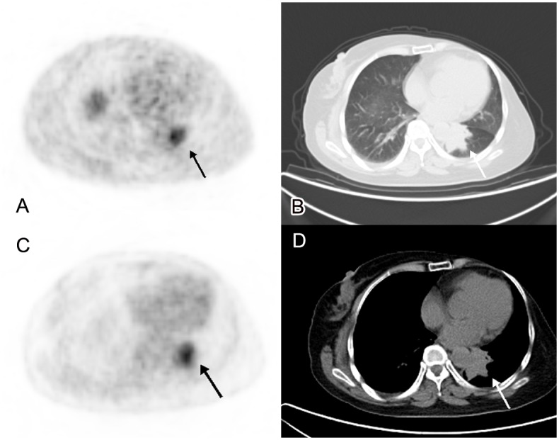
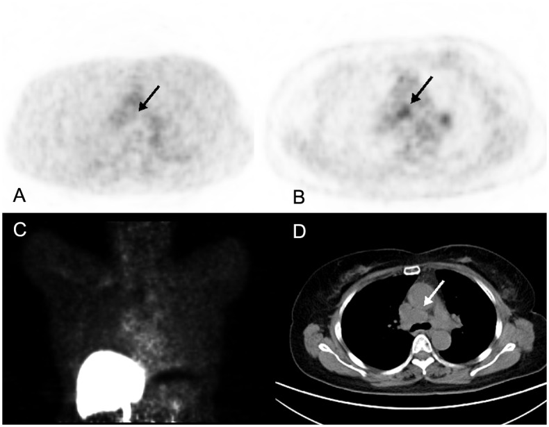
In evaluating equivocal lesions on conventional work-up group (n = 4), three lung lesions and another iliac lesion were enrolled. As for three lung lesions, 18F-FES PET/CT showed one lesion with high uptake, which suggested it was an ER positive metastasis. The other two lesions were 18F-FES negative, which meant an ER negative metastasis or secondary primary tumor. Additionally, one iliac lesion was detected by MRI. 18F-FDG uptake was high at the suspected lesion, whereas 18F-FES uptake was absent; In predicting origin of metastasis group (n = 2), two breast cancer patients had secondary primary tumors were collected. They were 18F-FES negative, which showed low possibility of metastasis from breast cancer and they were all confirmed by biopsy. In detecting ER status in metastasis group (n = 27), 18F-FES PET/CT showed increased 18F-FES uptake in all metastatic lesions in 11 patients; absent in all lesions in 13 patients; and the remaining 3 patients had both 18F-FES positive and negative lesions. Totally, on the basis of the 18F-FES PET/CT results, we found changes in the treatment plans in 16 patients (48.5%, 16/33).
18F-FES PET/CT could assess the entire tumor volume receptor status; therefore, it may be used to assist the individualized treatment decisions of breast cancer patients.
评估16α-[18F]氟雌二醇(18F-FES)PET/CT在辅助乳腺癌患者个体化治疗决策中的临床价值。
本初步研究纳入了2010年7月至2013年3月期间在本中心同时接受18F-FES和18F-FDG PET/CT检查的33例乳腺癌患者。所有患者均将18F-FES PET/CT作为解决临床难题的诊断工具。我们使用最大标准化摄取值(SUVmax)来量化雌激素受体(ER)表达,并将1.5的临界值作为将结果分为ER阳性和阴性病变的标准。所有患者均接受了至少6个月的临床随访。
在评估传统检查结果不明确的病变组(n = 4)时,纳入了3个肺部病变和另1个髂骨病变。对于3个肺部病变,18F-FES PET/CT显示1个病变摄取高,提示为ER阳性转移。另外2个病变18F-FES阴性,意味着为ER阴性转移或第二原发性肿瘤。此外,通过磁共振成像(MRI)检测到1个髂骨病变。可疑病变处18F-FDG摄取高,而18F-FES摄取缺如;在预测转移来源组(n = 2)中,收集了2例患有第二原发性肿瘤的乳腺癌患者。它们18F-FES阴性,显示乳腺癌转移的可能性低,且均经活检证实。在检测转移组(n = 27)的ER状态时,18F-FES PET/CT显示11例患者所有转移病变中18F-FES摄取增加;13例患者所有病变中均无摄取;其余3例患者既有18F-FES阳性病变也有阴性病变。总体而言,基于18F-FES PET/CT结果,我们发现16例患者(48.5%,16/33)的治疗方案发生了改变。
18F-FES PET/CT可评估整个肿瘤体积的受体状态;因此,它可用于辅助乳腺癌患者的个体化治疗决策。