Institute of Cardiovascular Diseases of PLA, Xinqiao Hospital, Third Military Medical University, Chongqing, People's Republic of China.
PLoS One. 2012;7(9):e43922. doi: 10.1371/journal.pone.0043922. Epub 2012 Sep 11.
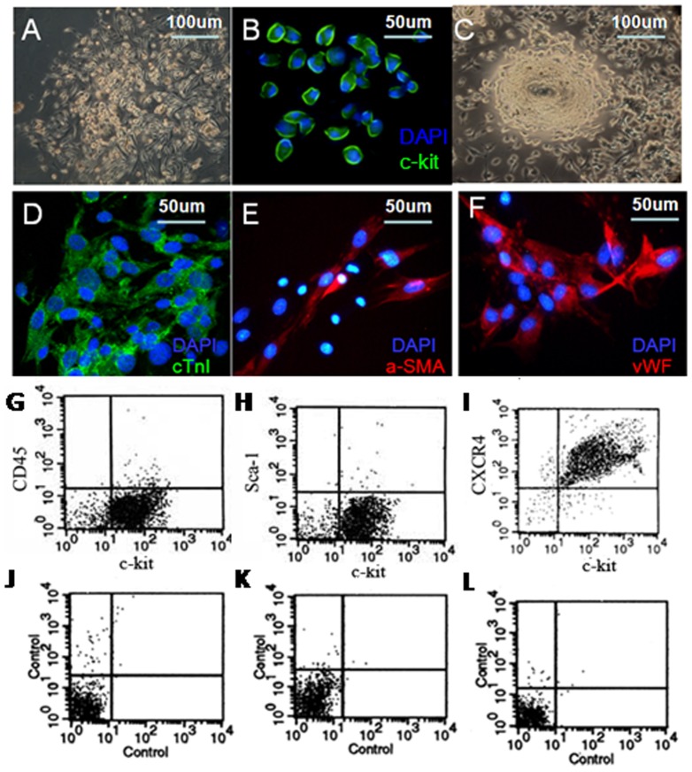
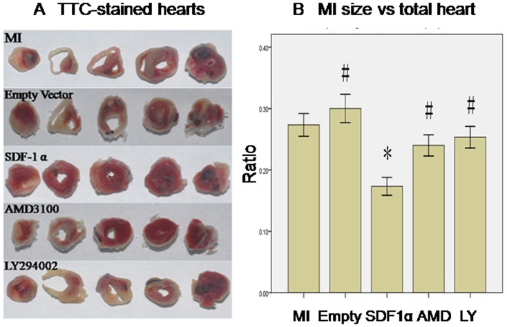
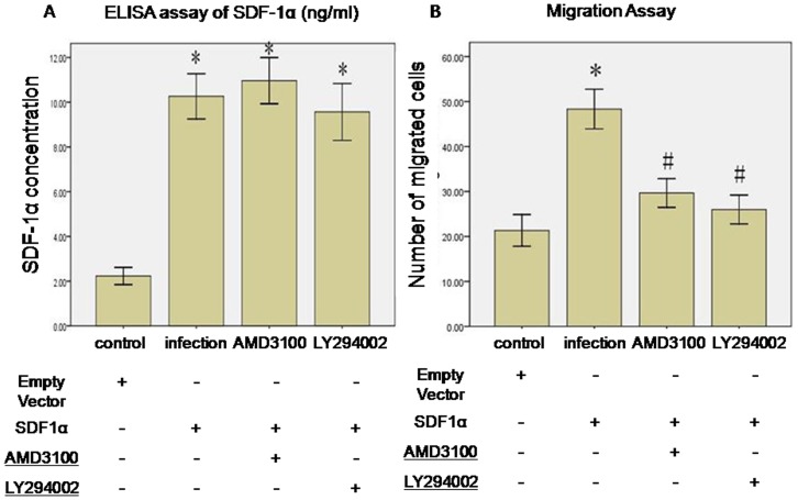
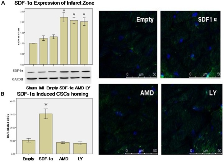
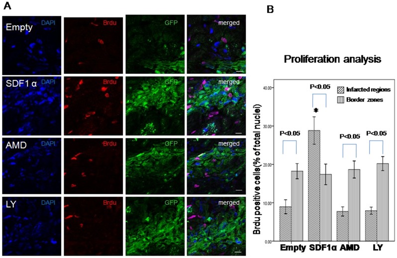
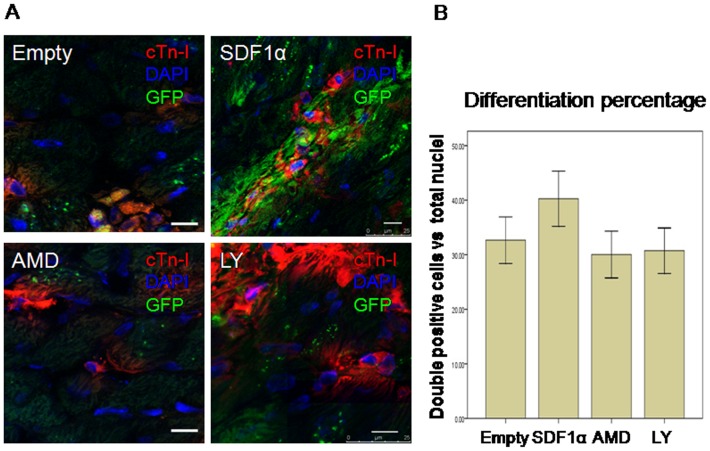
Cardiac stem cells (CSCs) can home to the infarcted area and regenerate myocardium. Stromal cell-derived factor-1α/C-X-C chemokine receptor type 4 (SDF-1α/CXCR4) axis is pivotal in inducing CSCs migration. However, the mechanisms remain unclear. This study set out to detect if SDF-1α promotes migration and engraftment of CSCs through the CXCR4/PI3K (phosphatidylinositol 3-kinase) pathway. In the in vitro experiment, c-kit+ cells were isolated from neonatal mouse heart fragment culture by magnetic cell sorting. Fluorescence-activated cell sorting results demonstrated that a few c-kit+ cells expressed CD45 (4.54%) and Sca-1 (2.58%), the hematopoietic stem cell marker. Conditioned culture could induce c-kit+ cells multipotent differentiation, which was confirmed by cardiac troponin I (cTn-I), α-smooth muscle actin (α-SMA), and von Willebrand factor (vWF) staining. In vitro chemotaxis assays were performed using Transwell cell chambers to detect CSCs migration. The results showed that the cardiomyocytes infected with rAAV1-SDF-1α-eGFP significantly increased SDF-1α concentration, 5-fold more in supernatant than that in the control group, and subsequently attracted more CSCs migration. This effect was diminished by administration of AMD3100 (10 µg/ml, CXCR4 antagonist) or LY294002 (20 µmol/L, PI3K inhibitor). In myocardial infarction mice, overexpression of SDF-1α in the infarcted area by rAAV1-SDF-1α-eGFP infection resulted in more CSCs retention to the infarcted myocardium, a higher percentage of proliferation, and reduced infarcted area which was attenuated by AMD3100 or ly294002 pretreatment. These results indicated that overexpression of SDF-1α enhanced CSCs migration in vitro and engraftment of transplanted CSCs and reduced infarcted size via CXCR4/PI3K pathway.
心脏干细胞 (CSCs) 可以归巢到梗死区域并再生心肌。基质细胞衍生因子-1α/C-X-C 趋化因子受体 4 (SDF-1α/CXCR4) 轴在诱导 CSCs 迁移中起着关键作用。然而,其机制尚不清楚。本研究旨在检测 SDF-1α 是否通过 CXCR4/PI3K(磷脂酰肌醇 3-激酶)途径促进 CSCs 的迁移和植入。在体外实验中,通过磁细胞分选从新生鼠心脏片段培养物中分离 c-kit+细胞。流式细胞术结果表明,少数 c-kit+细胞表达造血干细胞标志物 CD45(4.54%)和 Sca-1(2.58%)。条件培养可以诱导 c-kit+细胞多能分化,这通过心肌肌钙蛋白 I(cTn-I)、α-平滑肌肌动蛋白(α-SMA)和血管性血友病因子(vWF)染色得到证实。通过 Transwell 细胞室进行体外趋化实验来检测 CSCs 迁移。结果表明,感染 rAAV1-SDF-1α-eGFP 的心肌细胞显著增加了 SDF-1α 浓度,上清液中的浓度是对照组的 5 倍,随后吸引了更多的 CSCs 迁移。该作用被 AMD3100(10 µg/ml,CXCR4 拮抗剂)或 LY294002(20 µmol/L,PI3K 抑制剂)给药减弱。在心肌梗死小鼠中,rAAV1-SDF-1α-eGFP 感染使 SDF-1α 在梗死区域的过表达导致更多的 CSCs 保留在梗死心肌中,增殖的比例更高,梗死面积减小,而 AMD3100 或 ly294002 预处理则减弱了这一作用。这些结果表明,SDF-1α 的过表达通过 CXCR4/PI3K 途径增强了 CSCs 的体外迁移和移植 CSCs 的植入,并减少了梗死面积。