Makaryus Amgad N, Henry Sonia, Loewinger Lee, Makaryus John N, Boxt Lawrence
North Shore-LIJ Health System, Hofstra NSLIJ School of Medicine, New York, USA. ; Department of Cardiology, NuHealth, Nassau University Medical Center, East Meadow, NY, USA.
North Shore-LIJ Health System, Hofstra NSLIJ School of Medicine, New York, USA.
Clin Med Insights Cardiol. 2015 Jan 5;8(Suppl 4):13-22. doi: 10.4137/CMC.S18223. eCollection 2014.
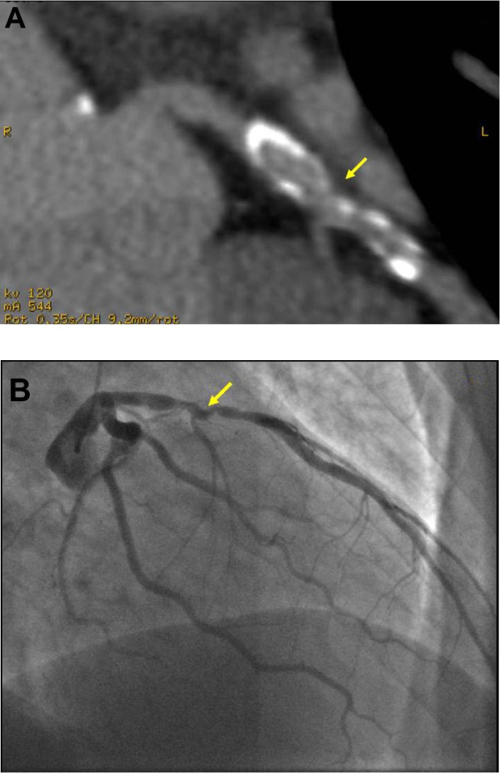
Multi-detector computed tomography (CT) has emerged as a modality for the non-invasive assessment of coronary artery disease (CAD). Prior studies have selected patients for evaluation and have excluded many of the "real-world" patients commonly encountered in daily practice. We compared 64-detector-CT (64-CT) to conventional coronary angiography (CA) to investigate the accuracy of 64-CT in determining significant coronary stenoses in a "real-world" clinical population.
A total of 1,818 consecutive patients referred for 64-CT were evaluated. CT angiography was performed using the GE LightSpeed VCT (GE(®) Healthcare). Forty-one patients in whom 64-CT results prompted CA investigation were further evaluated, and results of the two diagnostic modalities were compared.
A total of 164 coronary arteries and 410 coronary segments were evaluated in 41 patients (30 men, 11 women, age 39-85 years) who were identified by 64-CT to have significant coronary stenoses and who thereafter underwent CA. The overall per-vessel sensitivity, specificity, positive predictive value, negative predictive value, and accuracy at the 50% stenosis level were 86%, 84%, 65%, 95%, and 85%, respectively, and 77%, 93%, 61%, 97%, and 91%, respectively, in the per-segment analysis at the 50% stenosis level.
64-CT is an accurate imaging tool that allows a non-invasive assessment of significant CAD with a high diagnostic accuracy in a "real-world" population of patients. The sensitivity and specificity that we noted are not as high as those in prior reports, but we evaluated a population of patients that is typically encountered in clinical practice and therefore see more "real-world" results.
多排螺旋计算机断层扫描(CT)已成为一种用于冠状动脉疾病(CAD)无创评估的方式。先前的研究选择患者进行评估,并排除了日常临床实践中常见的许多“真实世界”患者。我们将64排CT(64-CT)与传统冠状动脉造影(CA)进行比较,以研究64-CT在“真实世界”临床人群中确定显著冠状动脉狭窄的准确性。
对连续1818例接受64-CT检查的患者进行评估。使用GE LightSpeed VCT(GE(®)医疗保健公司)进行CT血管造影。对64-CT结果提示进行CA检查的41例患者进行进一步评估,并比较两种诊断方式的结果。
在41例(30例男性,11例女性,年龄39-85岁)经64-CT确定有显著冠状动脉狭窄并随后接受CA检查的患者中,共评估了164条冠状动脉和410个冠状动脉节段。在50%狭窄水平的血管分析中,总体血管敏感性、特异性、阳性预测值、阴性预测值和准确性分别为86%、84%、65%、95%和85%;在节段分析中,50%狭窄水平时分别为77%、93%、61%、97%和91%。
64-CT是一种准确的成像工具,可在“真实世界”患者群体中对显著CAD进行无创评估,诊断准确性高。我们观察到的敏感性和特异性不如先前报告中的高,但我们评估的是临床实践中通常遇到的患者群体,因此看到了更多“真实世界”的结果。