Moradmand Hajar, Setayeshi Saeed, Karimian Ali Reza, Sirous Mehri, Akbari Mohammad Esmaeil
Dept. of Biomedical Radiation Engineering, Amirkabir University of Technology, Tehran, Iran.
Dept. of Biomedical Engineering, Faculty of Engineering, University of Isfahan, Isfahan, Iran.
Iran J Cancer Prev. 2012 Spring;5(2):61-8.
Mammography is the primary imaging technique for detection and diagnosis of breast cancer; however, the contrast of a mammogram image is often poor, especially for dense and glandular tissues. In these cases the radiologist may miss some diagnostically important microcalcifications. In order to improve diagnosis of cancer correctly, image enhancement technology is often used to enhance the image and help radiologists.
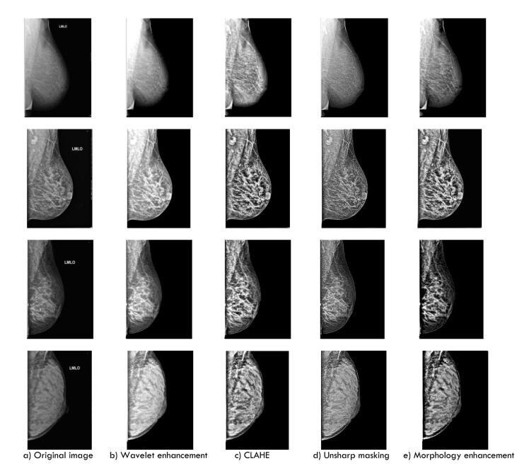
This paper presents a comparative study in digital mammography image enhancement based on four different algorithms: wavelet-based enhancement (Asymmetric Daubechies of order 8), Contrast-Limited Adaptive Histogram Equalization (CLAHE), morphological operators and unsharp masking. These algorithms have been tested on 114 clinical digital mammography images. The comparison for all the proposed image enhancement techniques was carried out to find out the best technique in enhancement of the mammogram images to detect microcalcifications.
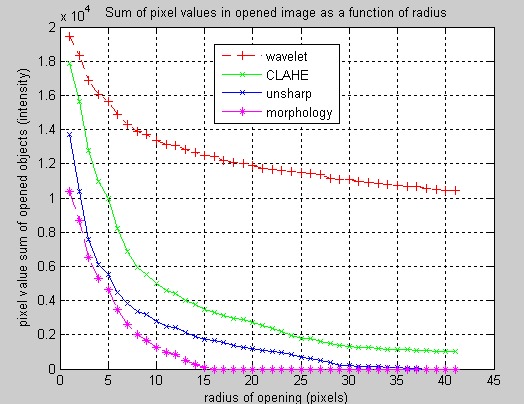
For evaluation of performance of image enhancement algorithms, the Contrast Improvement Index (CII) and profile intensity surface area distribution curve quality assessment have been used after any enhancement. The results of this study have shown that the average of CII is about 2.61 for wavelet and for CLAHE, unsharp masking and morphology operation are about 2.047, 1.63 and 1.315 respectively.
Experimental results strongly suggest that the wavelet transformation can be more effective and improve significantly overall detection of the Computer-Aided Diagnosis (CAD) system especially for dense breast. Compare to other studies, our method achieved a higher CII.
乳腺钼靶摄影是检测和诊断乳腺癌的主要成像技术;然而,乳腺钼靶图像的对比度通常较差,尤其是对于致密和腺性组织。在这些情况下,放射科医生可能会遗漏一些具有诊断重要性的微钙化。为了正确改善癌症诊断,图像增强技术经常被用于增强图像并帮助放射科医生。
本文基于四种不同算法对数字乳腺钼靶图像增强进行了对比研究:基于小波的增强(8阶非对称Daubechies小波)、对比度受限自适应直方图均衡化(CLAHE)、形态学算子和锐化掩模。这些算法已在114幅临床数字乳腺钼靶图像上进行了测试。对所有提出的图像增强技术进行比较,以找出在增强乳腺钼靶图像以检测微钙化方面的最佳技术。
为了评估图像增强算法的性能,在任何增强后都使用了对比度改善指数(CII)和轮廓强度表面积分布曲线质量评估。本研究结果表明,小波变换的CII平均值约为2.61,CLAHE、锐化掩模和形态学运算的CII平均值分别约为2.047、1.63和1.315。
实验结果强烈表明,小波变换可能更有效,并且能显著改善计算机辅助诊断(CAD)系统的整体检测效果,尤其是对于致密型乳腺。与其他研究相比,我们的方法获得了更高的CII。