Mouchtouris Nikolaos, Chalouhi Nohra, Chitale Ameet, Starke Robert M, Tjoumakaris Stavropoula I, Rosenwasser Robert H, Jabbour Pascal M
Department of Neurological Surgery, Thomas Jefferson University and Jefferson Hospital for Neuroscience, Philadelphia, PA 19107, USA.
Department of Neurological Surgery, University of Virginia School of Medicine, Charlottesville, VA 22908, USA.
ScientificWorldJournal. 2015;2015:808314. doi: 10.1155/2015/808314. Epub 2015 Jan 5.
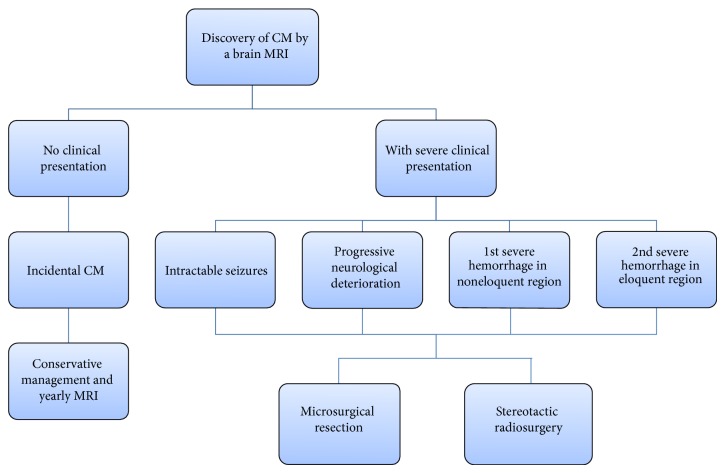
Cerebral cavernous malformations are the most common vascular malformations and can be found in many locations in the brain. If left untreated, cavernomas may lead to intracerebral hemorrhage, seizures, focal neurological deficits, or headaches. As they are angiographically occult, their diagnosis relies on various MR imaging techniques, which detect different characteristics of the lesions as well as aiding in planning the surgical treatment. The clinical presentation and the location of the lesion are the most important factors involved in determining the optimal course of treatment of cavernomas. We concisely review the literature and discuss the advantages and limitations of each of the three available methods of treatment--microsurgical resection, stereotactic radiosurgery, and conservative management--depending on the lesion characteristics.
脑海绵状血管畸形是最常见的血管畸形,可在脑内多个部位发现。如果不进行治疗,海绵状血管瘤可能导致脑出血、癫痫发作、局灶性神经功能缺损或头痛。由于它们在血管造影上隐匿,其诊断依赖于各种磁共振成像技术,这些技术可检测病变的不同特征并有助于规划手术治疗。临床表现和病变位置是决定海绵状血管瘤最佳治疗方案的最重要因素。我们简要回顾文献,并根据病变特征讨论三种可用治疗方法——显微手术切除、立体定向放射外科和保守治疗——各自的优缺点。